
External Email - Use Caution
Hello Bruce,
Thanks for responding back. To your question RE: "what does the aseg look like in those cases? That is, is it the hippo/amygdala stuff failing or is the root cause further back in the stream (something that caused a bad aseg)"
In all 5 cases I mentioned, ASEG looks fine. In other words, ASEG masks for both Amygdala and Hippocampus cover the appropriate voxels relative to subjects' T1 space. However, in all cases (116 subjects I looked at) the latest subfield segmentation seem to *underestimate these regions, meaning ASEG always covers more voxels.
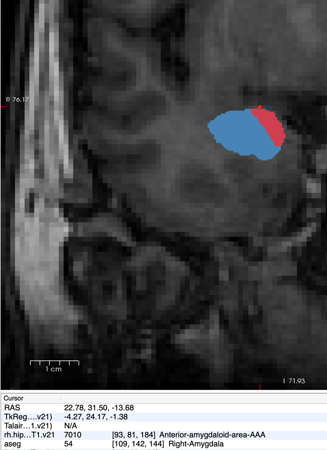
Along those lines, large portions of AAA regions are usually marked as "Cerebral Cortex" in ASEG. And the biggest issue again is that though AAA for 3 subjects are not visible in subfield segmentation mask, I get volumetric output. In one case *(Screenshot attached)*, R AAA has single voxel allocated, yet the volume is greater than the Left side which covers more space.
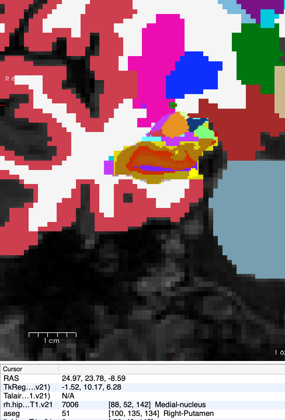
Also for the 2 cases in which medial nuclei are marked as Putamen *(Screenshot attached)*, ASEG of putamen and amygdala looks good, but the problematic voxels are darker compared to neighboring voxels *(Screenshot attached)*. This could be tissue damage and/or incorrect identification of "vessel".
What would be your recommendation to resolve these issues?
Thanks for your time,