Re: [Freesurfer] Reposting a Question RE: Hippocampal/Amygdala subfield segmentation - Anterior Amygdala Area / Medial Nucleus Errors

External Email - Use Caution
Hello Bruce,
Thanks for responding back. To your question RE: "what does the aseg look like in those cases? That is, is it the hippo/amygdala stuff failing or is the root cause further back in the stream (something that caused a bad aseg)"
In all 5 cases I mentioned, ASEG looks fine. In other words, ASEG masks for both Amygdala and Hippocampus cover the appropriate voxels relative to subjects' T1 space. However, in all cases (116 subjects I looked at) the latest subfield segmentation seem to *underestimate these regions, meaning ASEG always covers more voxels.
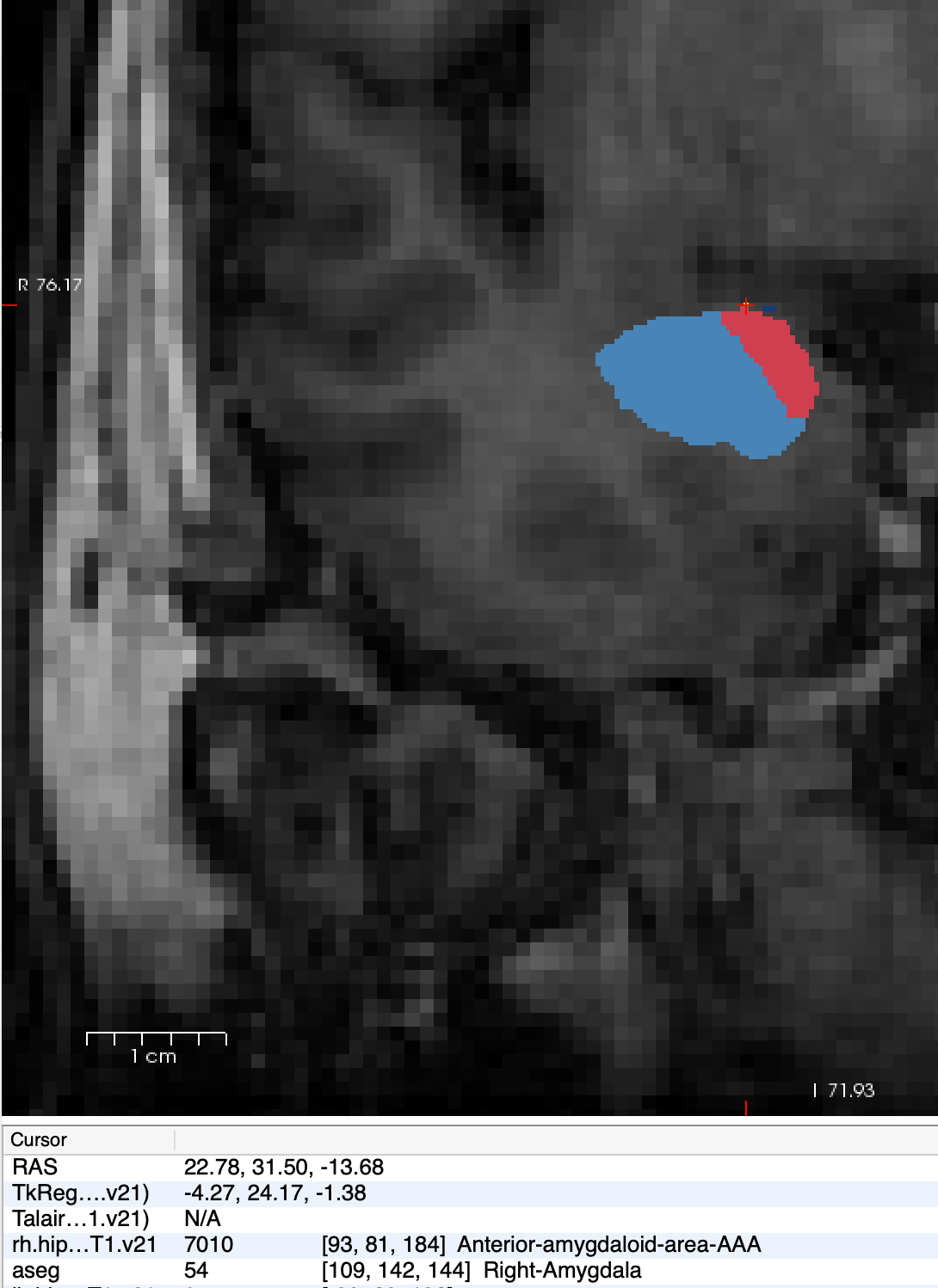
Along those lines, large portions of AAA regions are usually marked as "Cerebral Cortex" in ASEG. And the biggest issue again is that though AAA for 3 subjects are not visible in subfield segmentation mask, I get volumetric output. In one case *(Screenshot attached)*, R AAA has single voxel allocated, yet the volume is greater than the Left side which covers more space.
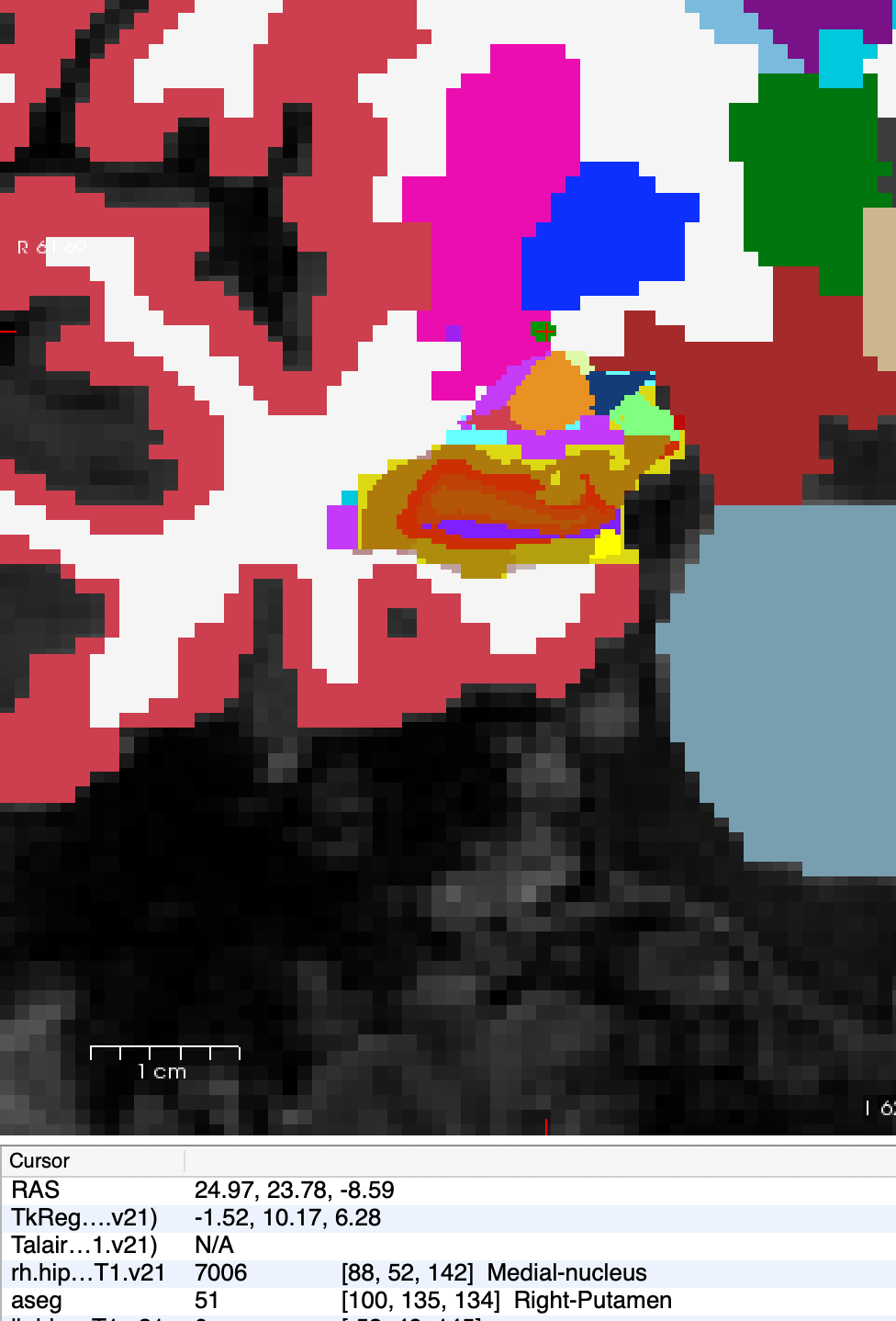
Also for the 2 cases in which medial nuclei are marked as Putamen *(Screenshot attached)*, ASEG of putamen and amygdala looks good, but the problematic voxels are darker compared to neighboring voxels *(Screenshot attached)*. This could be tissue damage and/or incorrect identification of "vessel".
What would be your recommendation to resolve these issues?
Thanks for your time,

External Email - Use Caution
Dear David,
These are all great questions.
ASEG covers more voxels: the segmentations from the HA module are leaner than their ASEG counterparts (the volumes are very highly correlated, though). This is because the HA module operates at higher resolution, and also because an algorithm is expected to have more false positives when you need to label *every voxel* as something.
Large portions of AAA are marked as cerebral cortex: when building the amygdala atlas, our anatomy experts noticed that parts of the amygdala were labelled as cortex by ASEG. This was corrected in the HA module, which we believe is more accurate in that region.
Mismatch between segmentations and volumes: these can happen because the volume is not computed with the hard segmentations (i.e., simply counting voxels), but using soft segmentations instead. So, there are voxels that may contribute to the volume but do not appear in the segmentation because another label was more likely at that location.
I hope this helps.
/Eugenio
-- Juan Eugenio Iglesias Senior research fellow CMIC (UCL), MGH (HMS) and CSAIL (MIT) http://www.jeiglesias.com
From: freesurfer-bounces@nmr.mgh.harvard.edu on behalf of David S Lee david.s.lee@wisc.edu Reply-To: Freesurfer support list freesurfer@nmr.mgh.harvard.edu Date: Tuesday, 25 June 2019 at 15:03 To: "freesurfer@nmr.mgh.harvard.edu" freesurfer@nmr.mgh.harvard.edu Subject: Re: [Freesurfer] Reposting a Question RE: Hippocampal/Amygdala subfield segmentation - Anterior Amygdala Area / Medial Nucleus Errors
External Email - Use Caution Hello Bruce,
Thanks for responding back. To your question RE: "what does the aseg look like in those cases? That is, is it the hippo/amygdala stuff failing or is the root cause further back in the stream (something that caused a bad aseg)"
In all 5 cases I mentioned, ASEG looks fine. In other words, ASEG masks for both Amygdala and Hippocampus cover the appropriate voxels relative to subjects' T1 space. However, in all cases (116 subjects I looked at) the latest subfield segmentation seem to *underestimate these regions, meaning ASEG always covers more voxels.
Along those lines, large portions of AAA regions are usually marked as "Cerebral Cortex" in ASEG. And the biggest issue again is that though AAA for 3 subjects are not visible in subfield segmentation mask, I get volumetric output. In one case (Screenshot attached), R AAA has single voxel allocated, yet the volume is greater than the Left side which covers more space.
Also for the 2 cases in which medial nuclei are marked as Putamen (Screenshot attached), ASEG of putamen and amygdala looks good, but the problematic voxels are darker compared to neighboring voxels (Screenshot attached). This could be tissue damage and/or incorrect identification of "vessel".
What would be your recommendation to resolve these issues?
Thanks for your time,
-- David S. Lee Research Specialist Center for Healthy Minds University of Wisconsin - Madison (608) 890-1115tel:(608)%20890-1115
freesurfer@nmr.mgh.harvard.edu
-
 David S Lee
 -
 Iglesias Gonzalez, Eugenio