
15 May
2014
15 May
'14
12:59 p.m.
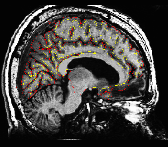
Hi all,
I'm having similar issues that were brought up in this post regarding subjects with cortical lesions: https://mail.nmr.mgh.harvard.edu/pipermail/freesurfer/2007-January/004359.ht...
Would these lesions would affect the way the brain is labeled and the measurements for cortical thickness? If so, how would this affect group comparisons of lesion patients to healthy controls?
I've attached an example of what one of the lesion subjects looks like.
Thanks, Maia