
Hi all,
I'm having similar issues that were brought up in this post regarding subjects with cortical lesions: https://mail.nmr.mgh.harvard.edu/pipermail/freesurfer/2007-January/004359.ht...
Would these lesions would affect the way the brain is labeled and the measurements for cortical thickness? If so, how would this affect group comparisons of lesion patients to healthy controls?
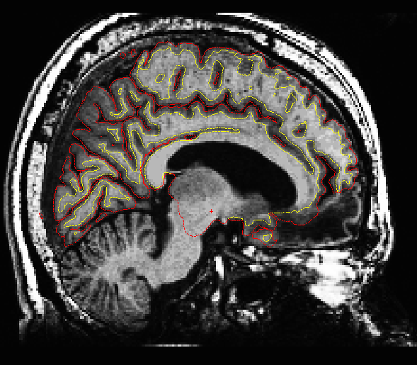
I've attached an example of what one of the lesion subjects looks like.
Thanks, Maia

On 05/15/2014 12:59 PM, Maia Pujara wrote:
Hi all,
I'm having similar issues that were brought up in this post regarding subjects with cortical lesions: https://mail.nmr.mgh.harvard.edu/pipermail/freesurfer/2007-January/004359.ht...
Would these lesions would affect the way the brain is labeled and the measurements for cortical thickness?
They certainly will in and near the lesions. Away from the lesions it will probably be ok.
If so, how would this affect group comparisons of lesion patients to healthy controls?
It depends on how big the lesion is and whether some cortical regions are not represented on the surface (as appears to be the case below). If so, then it could mess up the registration, possibly even far away from the lesion. Look at the ?h.aparc.annot to see if the boundaries look ok. If so, then I think it is safe to do the group analysis doug
I've attached an example of what one of the lesion subjects looks like.
Thanks, Maia
Freesurfer mailing list Freesurfer@nmr.mgh.harvard.edu https://mail.nmr.mgh.harvard.edu/mailman/listinfo/freesurfer
freesurfer@nmr.mgh.harvard.edu
-
 Douglas N Greve
 -
 Maia Pujara