
Hi team,
I am having problem with almost all the subjects in which wm/pial surfaces are not estimated correctly. These subjects are suffering from Corticobasal degeneration or progressive supranuclear palsy. I tried running sample of these subjects with multiple T1s as input (as we had them) and with additional parameters: -FLAIR, -FLAIRpial, -norm2-b 20, -norm2-n 5, -3T, and -bigventricles.
These are either mprage or spgr sequences with 1x1x1 resolution.
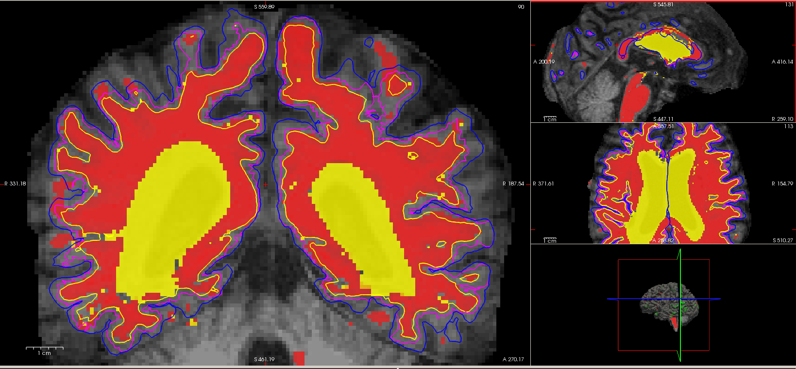
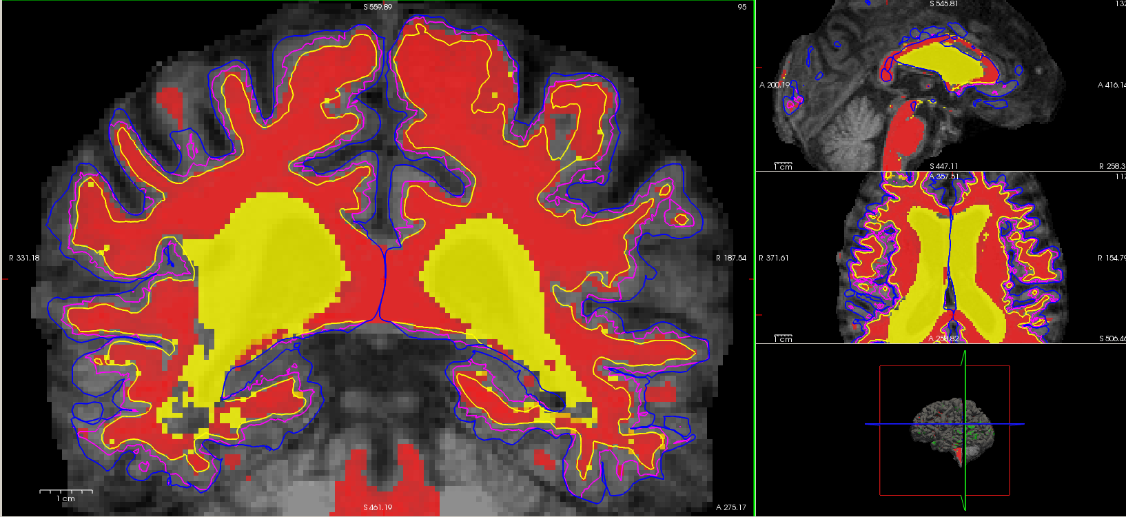
Flair input did not help instead made pial surfaces worst and convoluted (blue) as opposed to *.woFLAIR.pial surfaces (magenta), and even with addition of other flags it did not further improve estimation of wm/pial surfaces as seen in attached screenshots. The only option being adding control points. But these are about 200+ subjects, and I am not sure if to trust the manual edits on such a big scale.
Can anyone please suggest any other tweaks I could try?
[cid:01f72aae-1e98-4f12-bcf1-d2e3639dc992]
[cid:d1acff28-76d5-4cbf-aa2b-8fb55a07abfe]
Thanks, Sneha