
Dear all,
I've looked a little bit further into why LGI fails for fsaverage. I previously tried simply running recon-all -s fsaverage-localGI but the script exited with errors after a good while. More importantly for me, the lh.pial-outer-smoothed surface, which I am after, did not look right.
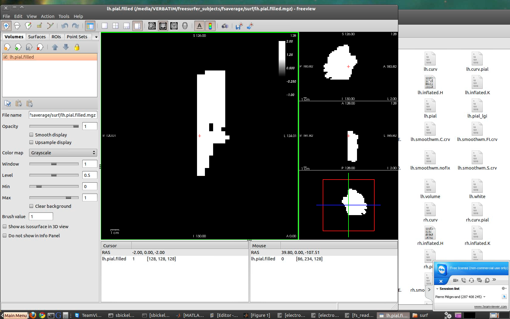
It seems that the first step, mris_fill, produces a volume that does not look very much like a brain anymore (cf. attached screenshot). Any idea why that would happen?
Thanks,
Pierre -- Pierre Mégevand, MD, PhD PLOS Neuro Community http://neuro.plos.org editor - Follow us on Twitter http://twitter.com/PLOSNeuro Postdoc @ Feinstein Institute for Medical Research (NY, USA) Follow me on Twitter http://twitter.com/pierre_vanmedge - Read my blog here http://neuroscimed.wordpress.com

Hi Pierre,
I'm not sure exactly what you are trying to do, but it's true that LGI fails for fsaverage at the mris_fills step, where the volume looks "cut". It seems to be something in the properties of the fsaverage surfaces. Maybe Doug or Bruce have an idea where it comes from.
But in any case, I wouldn't advice to run the LGI on the fsaverage, as the pattern of cortical folding is lost from the averaging of the subjects. So I'm not even sure that the algorithm will work even if you get mris_fill to work.
Can you tell us more precisely what you are trying to do? And why on the fsaverage rather than on your individual subject?
Best,
Marie
On Oct 28, 2014, at 5:31 AM, Pierre Mégevand <pierre.megevand@gmail.commailto:pierre.megevand@gmail.com> wrote:
Dear all,
I've looked a little bit further into why LGI fails for fsaverage. I previously tried simply running recon-all -s fsaverage-localGI but the script exited with errors after a good while. More importantly for me, the lh.pial-outer-smoothed surface, which I am after, did not look right.
It seems that the first step, mris_fill, produces a volume that does not look very much like a brain anymore (cf. attached screenshot). Any idea why that would happen?
Thanks,
Pierre -- Pierre Mégevand, MD, PhD PLOS Neuro Communityhttp://neuro.plos.org/ editor - Follow us on Twitterhttp://twitter.com/PLOSNeuro Postdoc @ Feinstein Institute for Medical Research (NY, USA) Follow me on Twitterhttp://twitter.com/pierre_vanmedge - Read my blog herehttp://neuroscimed.wordpress.com/ <screenshot3.png>_______________________________________________ Freesurfer mailing list Freesurfer@nmr.mgh.harvard.edumailto:Freesurfer@nmr.mgh.harvard.edu https://mail.nmr.mgh.harvard.edu/mailman/listinfo/freesurfer
The information in this e-mail is intended only for the person to whom it is addressed. If you believe this e-mail was sent to you in error and the e-mail contains patient information, please contact the Partners Compliance HelpLine at http://www.partners.org/complianceline . If the e-mail was sent to you in error but does not contain patient information, please contact the sender and properly dispose of the e-mail.

It does not surprise me that it fails on the fsaverage surface. I agree with Marie that it is not good idea to use LGI on fsaverage for anything. Definitely not its intended purpose. doug
On 10/28/2014 03:06 PM, Marie Schaer wrote:
Hi Pierre,
I'm not sure exactly what you are trying to do, but it's true that LGI fails for fsaverage at the mris_fills step, where the volume looks "cut". It seems to be something in the properties of the fsaverage surfaces. Maybe Doug or Bruce have an idea where it comes from.
But in any case, I wouldn't advice to run the LGI on the fsaverage, as the pattern of cortical folding is lost from the averaging of the subjects. So I'm not even sure that the algorithm will work even if you get mris_fill to work.
Can you tell us more precisely what you are trying to do? And why on the fsaverage rather than on your individual subject?
Best,
Marie
On Oct 28, 2014, at 5:31 AM, Pierre Mégevand <pierre.megevand@gmail.com mailto:pierre.megevand@gmail.com> wrote:
Dear all,
I've looked a little bit further into why LGI fails for fsaverage. I previously tried simply running recon-all -s fsaverage-localGI but the script exited with errors after a good while. More importantly for me, the lh.pial-outer-smoothed surface, which I am after, did not look right.
It seems that the first step, mris_fill, produces a volume that does not look very much like a brain anymore (cf. attached screenshot). Any idea why that would happen?
Thanks,
Pierre
Pierre Mégevand, MD, PhD PLOS Neuro Community http://neuro.plos.org/ editor - Follow us on Twitter http://twitter.com/PLOSNeuro Postdoc @ Feinstein Institute for Medical Research (NY, USA) Follow me on Twitter http://twitter.com/pierre_vanmedge- Read my blog here http://neuroscimed.wordpress.com/ <screenshot3.png>_______________________________________________ Freesurfer mailing list Freesurfer@nmr.mgh.harvard.edu mailto:Freesurfer@nmr.mgh.harvard.edu https://mail.nmr.mgh.harvard.edu/mailman/listinfo/freesurfer
The information in this e-mail is intended only for the person to whom it is addressed. If you believe this e-mail was sent to you in error and the e-mail contains patient information, please contact the Partners Compliance HelpLine at http://www.partners.org/complianceline . If the e-mail was sent to you in error but does not contain patient information, please contact the sender and properly dispose of the e-mail.
Freesurfer mailing list Freesurfer@nmr.mgh.harvard.edu https://mail.nmr.mgh.harvard.edu/mailman/listinfo/freesurfer
Thanks Marie. Here is what I'm trying to do. In fact, all I need from LGI are the first few steps, until ?h.pial-outer-smoothed has been generated. I'm not actually interested in the local GI for fsaverage.
Our intra-cranial electrode localization method relies on post-implant CT and MRI scans to localize the electrodes, and a pre-implant MR scan to which we co-register the post-op exams and to whose smoothed outer pial surface we then "snap" the electrodes to, in order to account for the brain shift caused by the implantation procedure (Dykstra et al., 2012).
Now, for one of our patients, the pre-op MRI was acquired with a lot of gadolinium, and we can't get Freesurfer to compute the pial surface. So I thought I would co-register the post-op exams to the fsaverage brain, and then snap the electrodes to fsaverage's outer smoothed pial surface as an approximation. Any idea how else I could do this?
Thanks,
Pierre On Oct 28, 2014 3:06 PM, "Marie Schaer" Marie.Schaer@unige.ch wrote:
Hi Pierre,
I'm not sure exactly what you are trying to do, but it's true that LGI fails for fsaverage at the mris_fills step, where the volume looks "cut". It seems to be something in the properties of the fsaverage surfaces. Maybe Doug or Bruce have an idea where it comes from.
But in any case, I wouldn't advice to run the LGI on the fsaverage, as the pattern of cortical folding is lost from the averaging of the subjects. So I'm not even sure that the algorithm will work even if you get mris_fill to work.
Can you tell us more precisely what you are trying to do? And why on the fsaverage rather than on your individual subject?
Best,
Marie
On Oct 28, 2014, at 5:31 AM, Pierre Mégevand pierre.megevand@gmail.com wrote:
Dear all,
I've looked a little bit further into why LGI fails for fsaverage. I previously tried simply running recon-all -s fsaverage-localGI but the script exited with errors after a good while. More importantly for me, the lh.pial-outer-smoothed surface, which I am after, did not look right.
It seems that the first step, mris_fill, produces a volume that does not look very much like a brain anymore (cf. attached screenshot). Any idea why that would happen?
Thanks,
Pierre
Pierre Mégevand, MD, PhD PLOS Neuro Community http://neuro.plos.org/ editor - Follow us on Twitter http://twitter.com/PLOSNeuro Postdoc @ Feinstein Institute for Medical Research (NY, USA) Follow me on Twitter http://twitter.com/pierre_vanmedge - Read my blog here http://neuroscimed.wordpress.com/ <screenshot3.png>_______________________________________________ Freesurfer mailing list Freesurfer@nmr.mgh.harvard.edu https://mail.nmr.mgh.harvard.edu/mailman/listinfo/freesurfer
The information in this e-mail is intended only for the person to whom it is addressed. If you believe this e-mail was sent to you in error and the e-mail contains patient information, please contact the Partners Compliance HelpLine at http://www.partners.org/complianceline . If the e-mail was sent to you in error but does not contain patient information, please contact the sender and properly dispose of the e-mail.
Hi Pierre,
Tricky question. So you can't really use the post-op cortical surface for this participant, right? Otherwise I would simply use the outer smoothed pial for this subject directly, which would be much more similar to the pre-op than the outer surface from the fsaverage (?). Otherwise, with the gadolinium MRI, maybe you can still get an adequate white surface, but I'm not sure that it would help you. The other option is to use the brain mask volume (which may work even with gadolinium if you are lucky, otherwise you can play with the watershed parameters), and tesselate a surface directly on this volume. But you'll get the two hemispheres at once (and probably the cerebellum as well), which may not be what you want?
Best,
Marie
On Oct 28, 2014, at 2:10 PM, Pierre Mégevand <pierre.megevand@gmail.commailto:pierre.megevand@gmail.com> wrote:
Thanks Marie. Here is what I'm trying to do. In fact, all I need from LGI are the first few steps, until ?h.pial-outer-smoothed has been generated. I'm not actually interested in the local GI for fsaverage.
Our intra-cranial electrode localization method relies on post-implant CT and MRI scans to localize the electrodes, and a pre-implant MR scan to which we co-register the post-op exams and to whose smoothed outer pial surface we then "snap" the electrodes to, in order to account for the brain shift caused by the implantation procedure (Dykstra et al., 2012).
Now, for one of our patients, the pre-op MRI was acquired with a lot of gadolinium, and we can't get Freesurfer to compute the pial surface. So I thought I would co-register the post-op exams to the fsaverage brain, and then snap the electrodes to fsaverage's outer smoothed pial surface as an approximation. Any idea how else I could do this?
Thanks,
Pierre
On Oct 28, 2014 3:06 PM, "Marie Schaer" <Marie.Schaer@unige.chmailto:Marie.Schaer@unige.ch> wrote:
Hi Pierre,
I'm not sure exactly what you are trying to do, but it's true that LGI fails for fsaverage at the mris_fills step, where the volume looks "cut". It seems to be something in the properties of the fsaverage surfaces. Maybe Doug or Bruce have an idea where it comes from.
But in any case, I wouldn't advice to run the LGI on the fsaverage, as the pattern of cortical folding is lost from the averaging of the subjects. So I'm not even sure that the algorithm will work even if you get mris_fill to work.
Can you tell us more precisely what you are trying to do? And why on the fsaverage rather than on your individual subject?
Best,
Marie
On Oct 28, 2014, at 5:31 AM, Pierre Mégevand <pierre.megevand@gmail.commailto:pierre.megevand@gmail.com> wrote:
Dear all,
I've looked a little bit further into why LGI fails for fsaverage. I previously tried simply running recon-all -s fsaverage-localGI but the script exited with errors after a good while. More importantly for me, the lh.pial-outer-smoothed surface, which I am after, did not look right.
It seems that the first step, mris_fill, produces a volume that does not look very much like a brain anymore (cf. attached screenshot). Any idea why that would happen?
Thanks,
Pierre -- Pierre Mégevand, MD, PhD PLOS Neuro Communityhttp://neuro.plos.org/ editor - Follow us on Twitterhttp://twitter.com/PLOSNeuro Postdoc @ Feinstein Institute for Medical Research (NY, USA) Follow me on Twitterhttp://twitter.com/pierre_vanmedge - Read my blog herehttp://neuroscimed.wordpress.com/ <screenshot3.png>_______________________________________________ Freesurfer mailing list Freesurfer@nmr.mgh.harvard.edumailto:Freesurfer@nmr.mgh.harvard.edu https://mail.nmr.mgh.harvard.edu/mailman/listinfo/freesurfer
The information in this e-mail is intended only for the person to whom it is addressed. If you believe this e-mail was sent to you in error and the e-mail contains patient information, please contact the Partners Compliance HelpLine at http://www.partners.org/complianceline . If the e-mail was sent to you in error but does not contain patient information, please contact the sender and properly dispose of the e-mail.
  You're right, we can't use the post-op surface because of
  the deformation caused by surgery and the electrode
  artifacts.
Getting a surface right from the brainmask would be OK, if we can't get anything better, but the nice thing about using the fsaverage surface is that you can plot and compare electrode locations from multiple patients. Just in case we have nothing better, though, how would you do it?
Thanks again!
-- Pierre Mégevand, MD, PhD PLOS Neuro Community http://neuro.plos.org editor - Follow us on Twitter http://twitter.com/PLOSNeuro Postdoc @ Feinstein Institute for Medical Research (NY, USA) Follow me on Twitter http://twitter.com/pierre_vanmedge - Read my blog here http://neuroscimed.wordpress.com
On Tue, Oct 28, 2014 at 5:23 PM, Marie Schaer Marie.Schaer@unige.ch wrote:
Hi Pierre,
Tricky question. So you can't really use the post-op cortical surface for this participant, right? Otherwise I would simply use the outer smoothed pial for this subject directly, which would be much more similar to the pre-op than the outer surface from the fsaverage (?). Otherwise, with the gadolinium MRI, maybe you can still get an adequate white surface, but I'm not sure that it would help you. The other option is to use the brain mask volume (which may work even with gadolinium if you are lucky, otherwise you can play with the watershed parameters), and tesselate a surface directly on this volume. But you'll get the two hemispheres at once (and probably the cerebellum as well), which may not be what you want?
Best,
Marie
On Oct 28, 2014, at 2:10 PM, Pierre Mégevand pierre.megevand@gmail.com wrote:
Thanks Marie. Here is what I'm trying to do. In fact, all I need from LGI are the first few steps, until ?h.pial-outer-smoothed has been generated. I'm not actually interested in the local GI for fsaverage.
Our intra-cranial electrode localization method relies on post-implant CT and MRI scans to localize the electrodes, and a pre-implant MR scan to which we co-register the post-op exams and to whose smoothed outer pial surface we then "snap" the electrodes to, in order to account for the brain shift caused by the implantation procedure (Dykstra et al., 2012).
Now, for one of our patients, the pre-op MRI was acquired with a lot of gadolinium, and we can't get Freesurfer to compute the pial surface. So I thought I would co-register the post-op exams to the fsaverage brain, and then snap the electrodes to fsaverage's outer smoothed pial surface as an approximation. Any idea how else I could do this?
Thanks,
Pierre On Oct 28, 2014 3:06 PM, "Marie Schaer" Marie.Schaer@unige.ch wrote:
Hi Pierre,
I'm not sure exactly what you are trying to do, but it's true that LGI fails for fsaverage at the mris_fills step, where the volume looks "cut". It seems to be something in the properties of the fsaverage surfaces. Maybe Doug or Bruce have an idea where it comes from.
But in any case, I wouldn't advice to run the LGI on the fsaverage, as the pattern of cortical folding is lost from the averaging of the subjects. So I'm not even sure that the algorithm will work even if you get mris_fill to work.
Can you tell us more precisely what you are trying to do? And why on the fsaverage rather than on your individual subject?
Best,
Marie
On Oct 28, 2014, at 5:31 AM, Pierre Mégevand pierre.megevand@gmail.com wrote:
Dear all,
I've looked a little bit further into why LGI fails for fsaverage. I previously tried simply running recon-all -s fsaverage-localGI but the script exited with errors after a good while. More importantly for me, the lh.pial-outer-smoothed surface, which I am after, did not look right.
It seems that the first step, mris_fill, produces a volume that does not look very much like a brain anymore (cf. attached screenshot). Any idea why that would happen?
Thanks,
Pierre
Pierre Mégevand, MD, PhD PLOS Neuro Community http://neuro.plos.org/ editor - Follow us on Twitter http://twitter.com/PLOSNeuro Postdoc @ Feinstein Institute for Medical Research (NY, USA) Follow me on Twitter http://twitter.com/pierre_vanmedge - Read my blog here http://neuroscimed.wordpress.com/ <screenshot3.png>_______________________________________________ Freesurfer mailing list Freesurfer@nmr.mgh.harvard.edu https://mail.nmr.mgh.harvard.edu/mailman/listinfo/freesurfer
The information in this e-mail is intended only for the person to whom it is addressed. If you believe this e-mail was sent to you in error and the e-mail contains patient information, please contact the Partners Compliance HelpLine at http://www.partners.org/complianceline . If the e-mail was sent to you in error but does not contain patient information, please contact the sender and properly dispose of the e-mail.
So what I don't get in your pipeline is how you'll register the post-op brain to fsaverage if you don't have any cortical surface in the post-op?
If I were you, if you want precise registration that includes the sulci, I'd go with the white surface from the pre-op gadolinium scan. And if you just want a rough registration of the skull shape, then you can probably find a way to create the outer surface directly on the gadolinium scan: if your brain mask is good, then mris_fill should work (maybe you need to binarize the data first (in any case a few lines of code in matlab should do the trick). If the brain mask is not good due to gadolinium, then either playing with mri_watershed (and maybe try mri_fill to separate the hemispheres / pons and cerebellum) and then mris_fill for the tessellation. Or if it doesn't work with mri_watershed, playing a bit with threshold with another skull-stripping program (e.g. MRIcro), then importing the volume back in FreeSurfer and use mris_fill to get a good tessellation. Both solutions will need you to play a bit, but I guess you cannot really afford to simply throw these data and take another subject!
Good luck,
Marie
On Oct 28, 2014, at 2:57 PM, Pierre Mégevand <pierre.megevand@gmail.commailto:pierre.megevand@gmail.com> wrote:
You're right, we can't use the post-op surface because of the deformation caused by surgery and the electrode artifacts.
Getting a surface right from the brainmask would be OK, if we can't get anything better, but the nice thing about using the fsaverage surface is that you can plot and compare electrode locations from multiple patients. Just in case we have nothing better, though, how would you do it?
Thanks again!
-- Pierre Mégevand, MD, PhD PLOS Neuro Communityhttp://neuro.plos.org/ editor - Follow us on Twitterhttp://twitter.com/PLOSNeuro Postdoc @ Feinstein Institute for Medical Research (NY, USA) Follow me on Twitterhttp://twitter.com/pierre_vanmedge - Read my blog herehttp://neuroscimed.wordpress.com/
On Tue, Oct 28, 2014 at 5:23 PM, Marie Schaer <Marie.Schaer@unige.chmailto:Marie.Schaer@unige.ch> wrote:
Hi Pierre,
Tricky question. So you can't really use the post-op cortical surface for this participant, right? Otherwise I would simply use the outer smoothed pial for this subject directly, which would be much more similar to the pre-op than the outer surface from the fsaverage (?). Otherwise, with the gadolinium MRI, maybe you can still get an adequate white surface, but I'm not sure that it would help you. The other option is to use the brain mask volume (which may work even with gadolinium if you are lucky, otherwise you can play with the watershed parameters), and tesselate a surface directly on this volume. But you'll get the two hemispheres at once (and probably the cerebellum as well), which may not be what you want?
Best,
Marie
On Oct 28, 2014, at 2:10 PM, Pierre Mégevand <pierre.megevand@gmail.commailto:pierre.megevand@gmail.com> wrote:
Thanks Marie. Here is what I'm trying to do. In fact, all I need from LGI are the first few steps, until ?h.pial-outer-smoothed has been generated. I'm not actually interested in the local GI for fsaverage.
Our intra-cranial electrode localization method relies on post-implant CT and MRI scans to localize the electrodes, and a pre-implant MR scan to which we co-register the post-op exams and to whose smoothed outer pial surface we then "snap" the electrodes to, in order to account for the brain shift caused by the implantation procedure (Dykstra et al., 2012).
Now, for one of our patients, the pre-op MRI was acquired with a lot of gadolinium, and we can't get Freesurfer to compute the pial surface. So I thought I would co-register the post-op exams to the fsaverage brain, and then snap the electrodes to fsaverage's outer smoothed pial surface as an approximation. Any idea how else I could do this?
Thanks,
Pierre
On Oct 28, 2014 3:06 PM, "Marie Schaer" <Marie.Schaer@unige.chmailto:Marie.Schaer@unige.ch> wrote:
Hi Pierre,
I'm not sure exactly what you are trying to do, but it's true that LGI fails for fsaverage at the mris_fills step, where the volume looks "cut". It seems to be something in the properties of the fsaverage surfaces. Maybe Doug or Bruce have an idea where it comes from.
But in any case, I wouldn't advice to run the LGI on the fsaverage, as the pattern of cortical folding is lost from the averaging of the subjects. So I'm not even sure that the algorithm will work even if you get mris_fill to work.
Can you tell us more precisely what you are trying to do? And why on the fsaverage rather than on your individual subject?
Best,
Marie
On Oct 28, 2014, at 5:31 AM, Pierre Mégevand <pierre.megevand@gmail.commailto:pierre.megevand@gmail.com> wrote:
Dear all,
I've looked a little bit further into why LGI fails for fsaverage. I previously tried simply running recon-all -s fsaverage-localGI but the script exited with errors after a good while. More importantly for me, the lh.pial-outer-smoothed surface, which I am after, did not look right.
It seems that the first step, mris_fill, produces a volume that does not look very much like a brain anymore (cf. attached screenshot). Any idea why that would happen?
Thanks,
Pierre -- Pierre Mégevand, MD, PhD PLOS Neuro Communityhttp://neuro.plos.org/ editor - Follow us on Twitterhttp://twitter.com/PLOSNeuro Postdoc @ Feinstein Institute for Medical Research (NY, USA) Follow me on Twitterhttp://twitter.com/pierre_vanmedge - Read my blog herehttp://neuroscimed.wordpress.com/ <screenshot3.png>_______________________________________________ Freesurfer mailing list Freesurfer@nmr.mgh.harvard.edumailto:Freesurfer@nmr.mgh.harvard.edu https://mail.nmr.mgh.harvard.edu/mailman/listinfo/freesurfer
The information in this e-mail is intended only for the person to whom it is addressed. If you believe this e-mail was sent to you in error and the e-mail contains patient information, please contact the Partners Compliance HelpLine at http://www.partners.org/complianceline . If the e-mail was sent to you in error but does not contain patient information, please contact the sender and properly dispose of the e-mail.
Our usual method for electrode localization remains in the patient's space: co-register post-op MR and CT with pre-op MR brainmask (our reference space) and snap electrodes to patient's outer pial surface.
My idea was: register the patient's (crappy) brainmask volume to the "Talairach" fsaverage volume (not surface), which I can correct manually; use the Talairach.xfm transform to bring the electrodes into that same fsaverage volume, and then snap the electrodes to fsaverage's outer pial surface. But that last step I can't perform. I'm aware that the localization would be pretty inaccurate.
I'll look into what you suggest Marie. In case all else fails, the last resort is to show the actual post-op MR and CT slices for electrodes of interest, but that just does not look as appealing as pial surfaces!
-- Pierre Mégevand, MD, PhD PLOS Neuro Community http://neuro.plos.org editor - Follow us on Twitter http://twitter.com/PLOSNeuro Postdoc @ Feinstein Institute for Medical Research (NY, USA) Follow me on Twitter http://twitter.com/pierre_vanmedge - Read my blog here http://neuroscimed.wordpress.com
On Wed, Oct 29, 2014 at 12:29 AM, Marie Schaer Marie.Schaer@unige.ch wrote:
So what I don't get in your pipeline is how you'll register the post-op brain to fsaverage if you don't have any cortical surface in the post-op?
If I were you, if you want precise registration that includes the sulci, I'd go with the white surface from the pre-op gadolinium scan. And if you just want a rough registration of the skull shape, then you can probably find a way to create the outer surface directly on the gadolinium scan: if your brain mask is good, then mris_fill should work (maybe you need to binarize the data first (in any case a few lines of code in matlab should do the trick). If the brain mask is not good due to gadolinium, then either playing with mri_watershed (and maybe try mri_fill to separate the hemispheres / pons and cerebellum) and then mris_fill for the tessellation. Or if it doesn't work with mri_watershed, playing a bit with threshold with another skull-stripping program (e.g. MRIcro), then importing the volume back in FreeSurfer and use mris_fill to get a good tessellation. Both solutions will need you to play a bit, but I guess you cannot really afford to simply throw these data and take another subject!
Good luck,
Marie
On Oct 28, 2014, at 2:57 PM, Pierre Mégevand pierre.megevand@gmail.com wrote:
You're right, we can't use the post-op surface because of the deformation caused by surgery and the electrode artifacts.
Getting a surface right from the brainmask would be OK, if we can't get anything better, but the nice thing about using the fsaverage surface is that you can plot and compare electrode locations from multiple patients. Just in case we have nothing better, though, how would you do it?
Thanks again!
-- Pierre Mégevand, MD, PhD PLOS Neuro Community http://neuro.plos.org/ editor - Follow us on Twitter http://twitter.com/PLOSNeuro Postdoc @ Feinstein Institute for Medical Research (NY, USA) Follow me on Twitter http://twitter.com/pierre_vanmedge - Read my blog here http://neuroscimed.wordpress.com/
On Tue, Oct 28, 2014 at 5:23 PM, Marie Schaer Marie.Schaer@unige.ch wrote:
Hi Pierre,
Tricky question. So you can't really use the post-op cortical surface for this participant, right? Otherwise I would simply use the outer smoothed pial for this subject directly, which would be much more similar to the pre-op than the outer surface from the fsaverage (?). Otherwise, with the gadolinium MRI, maybe you can still get an adequate white surface, but I'm not sure that it would help you. The other option is to use the brain mask volume (which may work even with gadolinium if you are lucky, otherwise you can play with the watershed parameters), and tesselate a surface directly on this volume. But you'll get the two hemispheres at once (and probably the cerebellum as well), which may not be what you want?
Best,
Marie
On Oct 28, 2014, at 2:10 PM, Pierre Mégevand pierre.megevand@gmail.com wrote:
Thanks Marie. Here is what I'm trying to do. In fact, all I need from LGI are the first few steps, until ?h.pial-outer-smoothed has been generated. I'm not actually interested in the local GI for fsaverage.
Our intra-cranial electrode localization method relies on post-implant CT and MRI scans to localize the electrodes, and a pre-implant MR scan to which we co-register the post-op exams and to whose smoothed outer pial surface we then "snap" the electrodes to, in order to account for the brain shift caused by the implantation procedure (Dykstra et al., 2012).
Now, for one of our patients, the pre-op MRI was acquired with a lot of gadolinium, and we can't get Freesurfer to compute the pial surface. So I thought I would co-register the post-op exams to the fsaverage brain, and then snap the electrodes to fsaverage's outer smoothed pial surface as an approximation. Any idea how else I could do this?
Thanks,
Pierre On Oct 28, 2014 3:06 PM, "Marie Schaer" Marie.Schaer@unige.ch wrote:
Hi Pierre,
I'm not sure exactly what you are trying to do, but it's true that LGI fails for fsaverage at the mris_fills step, where the volume looks "cut". It seems to be something in the properties of the fsaverage surfaces. Maybe Doug or Bruce have an idea where it comes from.
But in any case, I wouldn't advice to run the LGI on the fsaverage, as the pattern of cortical folding is lost from the averaging of the subjects. So I'm not even sure that the algorithm will work even if you get mris_fill to work.
Can you tell us more precisely what you are trying to do? And why on the fsaverage rather than on your individual subject?
Best,
Marie
On Oct 28, 2014, at 5:31 AM, Pierre Mégevand pierre.megevand@gmail.com wrote:
Dear all,
I've looked a little bit further into why LGI fails for fsaverage. I previously tried simply running recon-all -s fsaverage-localGI but the script exited with errors after a good while. More importantly for me, the lh.pial-outer-smoothed surface, which I am after, did not look right.
It seems that the first step, mris_fill, produces a volume that does not look very much like a brain anymore (cf. attached screenshot). Any idea why that would happen?
Thanks,
Pierre
Pierre Mégevand, MD, PhD PLOS Neuro Community http://neuro.plos.org/ editor - Follow us on Twitter http://twitter.com/PLOSNeuro Postdoc @ Feinstein Institute for Medical Research (NY, USA) Follow me on Twitter http://twitter.com/pierre_vanmedge - Read my blog here http://neuroscimed.wordpress.com/ <screenshot3.png>_______________________________________________ Freesurfer mailing list Freesurfer@nmr.mgh.harvard.edu https://mail.nmr.mgh.harvard.edu/mailman/listinfo/freesurfer
The information in this e-mail is intended only for the person to whom it is addressed. If you believe this e-mail was sent to you in error and the e-mail contains patient information, please contact the Partners Compliance HelpLine at http://www.partners.org/complianceline . If the e-mail was sent to you in error but does not contain patient information, please contact the sender and properly dispose of the e-mail.

have you tried using bbregister to register post-op MRI with the pre-op surfaces? On Wed, 29 Oct 2014, Pierre Mégevand wrote:
Our usual method for electrode localization remains in the patient's space: co-register post-op MR and CT with pre-op MR brainmask (our reference space) and snap electrodes to patient's outer pial surface.
My idea was: register the patient's (crappy) brainmask volume to the "Talairach" fsaverage volume (not surface), which I can correct manually; use the Talairach.xfm transform to bring the electrodes into that same fsaverage volume, and then snap the electrodes to fsaverage's outer pial surface. But that last step I can't perform. I'm aware that the localization would be pretty inaccurate. I'll look into what you suggest Marie. In case all else fails, the last resort is to show the actual post-op MR and CT slices for electrodes of interest, but that just does not look as appealing as pial surfaces!
-- Pierre Mégevand, MD, PhD PLOS Neuro Community editor - Follow us on Twitter Postdoc @ Feinstein Institute for Medical Research (NY, USA) Follow me on Twitter - Read my blog here
On Wed, Oct 29, 2014 at 12:29 AM, Marie Schaer Marie.Schaer@unige.ch wrote:
So what I don't get in your pipeline is how you'll register the post-op brain to fsaverage if you don't have any cortical surface in the post-op?
If I were you, if you want precise registration that includes the sulci, I'd go with the white surface from the pre-op gadolinium scan. And if you just want a rough registration of the skull shape, then you can probably find a way to create the outer surface directly on the gadolinium scan: if your brain mask is good, then mris_fill should work (maybe you need to binarize the data first (in any case a few lines of code in matlab should do the trick). If the brain mask is not good due to gadolinium, then either playing with mri_watershed (and maybe try mri_fill to separate the hemispheres / pons and cerebellum) and then mris_fill for the tessellation. Or if it doesn't work with mri_watershed, playing a bit with threshold with another skull-stripping program (e.g. MRIcro), then importing the volume back in FreeSurfer and use mris_fill to get a good tessellation. Both solutions will need you to play a bit, but I guess you cannot really afford to simply throw these data and take another subject!
Good luck,
Marie
On Oct 28, 2014, at 2:57 PM, Pierre Mégevand pierre.megevand@gmail.com wrote:
You're right, we can't use the post-op surface because of the deformation caused by surgery and the electrode artifacts.Getting a surface right from the brainmask would be OK, if we can't get anything better, but the nice thing about using the fsaverage surface is that you can plot and compare electrode locations from multiple patients. Just in case we have nothing better, though, how would you do it?
Thanks again!
-- Pierre Mégevand, MD, PhD PLOS Neuro Community editor - Follow us on Twitter Postdoc @ Feinstein Institute for Medical Research (NY, USA) Follow me on Twitter - Read my blog here
On Tue, Oct 28, 2014 at 5:23 PM, Marie Schaer Marie.Schaer@unige.ch wrote:
Hi Pierre,
Tricky question. So you can't really use the post-op cortical surface for this participant, right? Otherwise I would simply use the outer smoothed pial for this subject directly, which would be much more similar to the pre-op than the outer surface from the fsaverage (?). Otherwise, with the gadolinium MRI, maybe you can still get an adequate white surface, but I'm not sure that it would help you. The other option is to use the brain mask volume (which may work even with gadolinium if you are lucky, otherwise you can play with the watershed parameters), and tesselate a surface directly on this volume. But you'll get the two hemispheres at once (and probably the cerebellum as well), which may not be what you want?
Best,
Marie
On Oct 28, 2014, at 2:10 PM, Pierre Mégevand pierre.megevand@gmail.com wrote:
  Thanks Marie. Here is what I'm trying to do.
  In fact, all I need from LGI are the first few
  steps, until ?h.pial-outer-smoothed has been
  generated. I'm not actually interested in the
  local GI for fsaverage.

  Our intra-cranial electrode localization
  method relies on post-implant CT and MRI scans
  to localize the electrodes, and a pre-implant
  MR scan to which we co-register the post-op
  exams and to whose smoothed outer pial surface
  we then "snap" the electrodes to, in order to
  account for the brain shift caused by the
  implantation procedure (Dykstra et al., 2012).

  Now, for one of our patients, the pre-op MRI
  was acquired with a lot of gadolinium, and we
  can't get Freesurfer to compute the pial
  surface. So I thought I would co-register the
  post-op exams to the fsaverage brain, and then
  snap the electrodes to fsaverage's outer
  smoothed pial surface as an approximation. Any
  idea how else I could do this?

  Thanks,

  Pierre

  On Oct 28, 2014 3:06 PM, "Marie Schaer"
  <Marie.Schaer@unige.ch> wrote:
Hi Pierre,
I'm not sure exactly what you are trying to do, but it's true that LGI fails for fsaverage at the mris_fills step, where the volume looks "cut". It seems to be something in the properties of the fsaverage surfaces. Maybe Doug or Bruce have an idea where it comes from.
But in any case, I wouldn't advice to run the LGI on the fsaverage, as the pattern of cortical folding is lost from the averaging of the subjects. So I'm not even sure that the algorithm will work even if you get mris_fill to work.
Can you tell us more precisely what you are trying to do? And why on the fsaverage rather than on your individual subject?
Best,
Marie
On Oct 28, 2014, at 5:31 AM, Pierre Mégevand pierre.megevand@gmail.com wrote:
Dear all,I've looked a little bit further into why LGI fails for fsaverage. I previously tried simply running recon-all -s fsaverage-localGI but the script exited with errors after a good while. More importantly for me, the lh.pial-outer-smoothed surface, which I am after, did not look right.
It seems that the first step, mris_fill, produces a volume that does not look very much like a brain anymore (cf. attached screenshot). Any idea why that would happen?
Thanks,
Pierre
Pierre Mégevand, MD, PhD PLOS Neuro Community editor - Follow us on Twitter Postdoc @ Feinstein Institute for Medical Research (NY, USA) Follow me on Twitter - Read my blog here <screenshot3.png>_______________________________________________ Freesurfer mailing list Freesurfer@nmr.mgh.harvard.edu https://mail.nmr.mgh.harvard.edu/mailman/listinfo/freesurfer
The information in this e-mail is intended only for the person to whom it is addressed. If you believe this e-mail was sent to you in error and the e-mail contains patient information, please contact the Partners Compliance HelpLine at http://www.partners.org/complianceline . If the e-mail was sent to you in error but does not contain patient information, please contact the sender and properly dispose of the e-mail.
freesurfer@nmr.mgh.harvard.edu
-
 Bruce Fischl
 -
 Douglas N Greve
 -
 Marie Schaer
 -
 Pierre Mégevand