
External Email - Use Caution
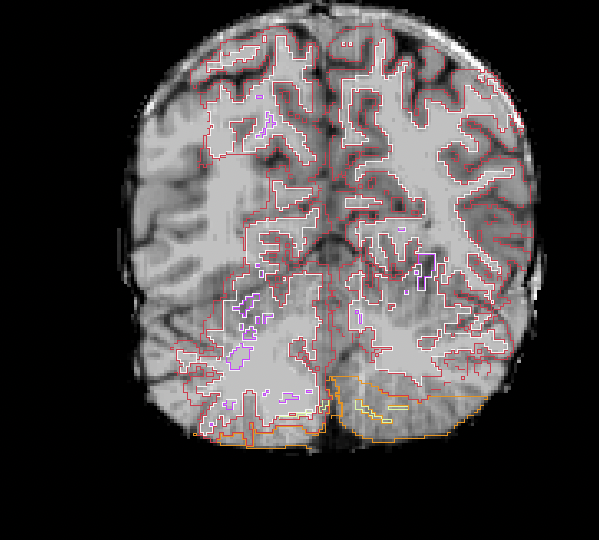
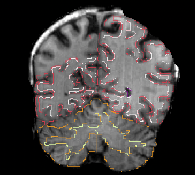
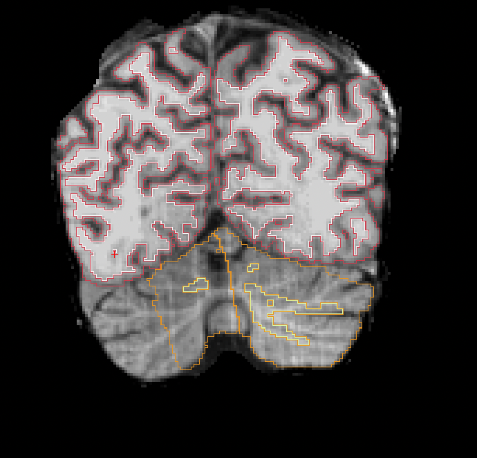
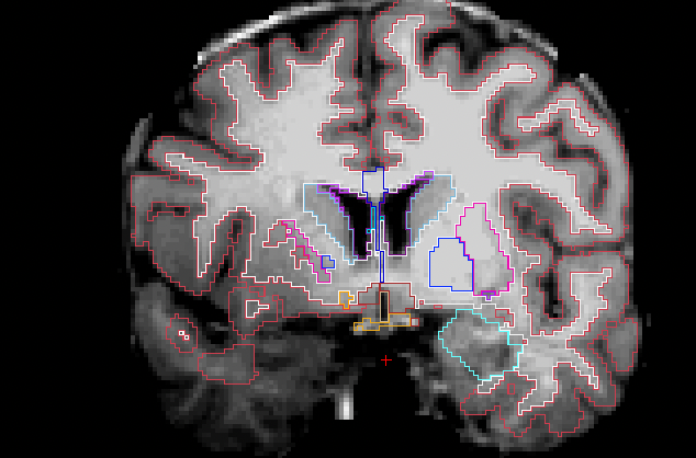
Dear FS, I intend to examine cortical thickness correlates of negative symptoms in my schizophrenia sample. I ran recon-all and corrected a few brainmasks which were excessively stripped by watershedding with different h values (ran autorecon-pial for those watershedded). I am attaching a few screenshots of scans where the segmentation is erroneous and the aseg omits the right temporal and parietal or cerebellar regions. How do I set this right?
[image: Screenshot 2023-01-27 at 3.37.46 PM.png] [image: Screenshot 2023-01-27 at 3.29.47 PM.png][image: Screenshot 2023-01-27 at 3.26.29 PM.png][image: Screenshot 2023-01-27 at 3.32.08 PM.png]
Dr. Rakshathi Basavaraju (M.D. Psychiatry) Assistant Professor (Department of Health Research-sponsored return-home research fellowship) Principal Investigator - OPCINSS Department of Psychiatry National Institute of Mental Health and Neurosciences (NIMHANS) Bengaluru, Karnataka, India-560029 Phone: +91-9480829898/+91-80-26972156/+91-80-26995320
External Email - Use Caution
Reposting this question.
Dr. Rakshathi Basavaraju (M.D. Psychiatry) Assistant Professor (Department of Health Research-sponsored return-home research fellowship) Principal Investigator - OPCINSS Department of Psychiatry National Institute of Mental Health and Neurosciences (NIMHANS) Bengaluru, Karnataka, India-560029 Phone: +91-9480829898/+91-80-26972156/+91-80-26995320
---------- Forwarded message --------- From: Rakshathi Basavaraju rakshathi.b@gmail.com Date: Fri, Jan 27, 2023 at 3:46 PM Subject: A doubt regarding autorecon after watershedding To: Freesurfer@nmr.mgh.harvard.edu
Dear FS, I intend to examine cortical thickness correlates of negative symptoms in my schizophrenia sample. I ran recon-all and corrected a few brainmasks which were excessively stripped by watershedding with different h values (ran autorecon-pial for those watershedded). I am attaching a few screenshots of scans where the segmentation is erroneous and the aseg omits the right temporal and parietal or cerebellar regions. How do I set this right?
[image: Screenshot 2023-01-27 at 3.37.46 PM.png] [image: Screenshot 2023-01-27 at 3.29.47 PM.png][image: Screenshot 2023-01-27 at 3.26.29 PM.png][image: Screenshot 2023-01-27 at 3.32.08 PM.png]
Dr. Rakshathi Basavaraju (M.D. Psychiatry) Assistant Professor (Department of Health Research-sponsored return-home research fellowship) Principal Investigator - OPCINSS Department of Psychiatry National Institute of Mental Health and Neurosciences (NIMHANS) Bengaluru, Karnataka, India-560029 Phone: +91-9480829898/+91-80-26972156/+91-80-26995320

Hi Again
You need to look through the processing steps and see where things went wrong. Is this a watershed failure? Or a topology fixing one? If watershed you can try mri_synthstrip and see if it fixes your problem. If topology correction you need to figure out where it is getting hung up Cheers Bruce
From: freesurfer-bounces@nmr.mgh.harvard.edu freesurfer-bounces@nmr.mgh.harvard.edu On Behalf Of Rakshathi Basavaraju Sent: Saturday, January 28, 2023 4:20 AM To: Freesurfer@nmr.mgh.harvard.edu Subject: [Freesurfer] Fwd: A doubt regarding autorecon after watershedding
External Email - Use Caution Reposting this question.
Dr. Rakshathi Basavaraju (M.D. Psychiatry) Assistant Professor (Department of Health Research-sponsored return-home research fellowship) Principal Investigator - OPCINSS Department of Psychiatry National Institute of Mental Health and Neurosciences (NIMHANS) Bengaluru, Karnataka, India-560029 Phone: +91-9480829898/+91-80-26972156/+91-80-26995320
---------- Forwarded message --------- From: Rakshathi Basavaraju <rakshathi.b@gmail.commailto:rakshathi.b@gmail.com> Date: Fri, Jan 27, 2023 at 3:46 PM Subject: A doubt regarding autorecon after watershedding To: <Freesurfer@nmr.mgh.harvard.edumailto:Freesurfer@nmr.mgh.harvard.edu>
Dear FS, I intend to examine cortical thickness correlates of negative symptoms in my schizophrenia sample. I ran recon-all and corrected a few brainmasks which were excessively stripped by watershedding with different h values (ran autorecon-pial for those watershedded). I am attaching a few screenshots of scans where the segmentation is erroneous and the aseg omits the right temporal and parietal or cerebellar regions. How do I set this right?
[cid:image001.png@01D9330A.883EB200] [cid:image002.png@01D9330A.883EB200][cid:image003.png@01D9330A.883EB200][cid:image004.png@01D9330A.883EB200]
Dr. Rakshathi Basavaraju (M.D. Psychiatry) Assistant Professor (Department of Health Research-sponsored return-home research fellowship) Principal Investigator - OPCINSS Department of Psychiatry National Institute of Mental Health and Neurosciences (NIMHANS) Bengaluru, Karnataka, India-560029 Phone: +91-9480829898/+91-80-26972156/+91-80-26995320
External Email - Use Caution
Hi,
Reposting this message as I do not see this in the FS archives. Any help would be appreciated.
Thanks, Dr. Rakshathi Basavaraju (M.D. Psychiatry) Assistant Professor (Department of Health Research-sponsored return-home research fellowship) Principal Investigator - OPCINSS Department of Psychiatry National Institute of Mental Health and Neurosciences (NIMHANS) Bengaluru, Karnataka, India-560029 Phone: +91-9480829898/+91-80-26972156/+91-80-26995320
---------- Forwarded message --------- From: Rakshathi Basavaraju rakshathi.b@gmail.com Date: Fri, Jan 27, 2023 at 3:46 PM Subject: A doubt regarding autorecon after watershedding To: Freesurfer@nmr.mgh.harvard.edu
Dear FS, I intend to examine cortical thickness correlates of negative symptoms in my schizophrenia sample. I ran recon-all and corrected a few brainmasks which were excessively stripped by watershedding with different h values (ran autorecon-pial for those watershedded). I am attaching a few screenshots of scans where the segmentation is erroneous and the aseg omits the right temporal and parietal or cerebellar regions. How do I set this right?
[image: Screenshot 2023-01-27 at 3.37.46 PM.png] [image: Screenshot 2023-01-27 at 3.29.47 PM.png][image: Screenshot 2023-01-27 at 3.26.29 PM.png][image: Screenshot 2023-01-27 at 3.32.08 PM.png]
Dr. Rakshathi Basavaraju (M.D. Psychiatry) Assistant Professor (Department of Health Research-sponsored return-home research fellowship) Principal Investigator - OPCINSS Department of Psychiatry National Institute of Mental Health and Neurosciences (NIMHANS) Bengaluru, Karnataka, India-560029 Phone: +91-9480829898/+91-80-26972156/+91-80-26995320
Looks like the talairach auto detection thinks it failed. The most common cause for this is when your initial volume has incorrect direction cosines. Can you bring up the input volume in freeview and verify that the direction freeview things are e.g. left/right are in fact left/right in the brain? If they are you should check out tutorial on fixing a bad tal xform
From: freesurfer-bounces@nmr.mgh.harvard.edu freesurfer-bounces@nmr.mgh.harvard.edu On Behalf Of Rakshathi Basavaraju Sent: Monday, January 30, 2023 2:24 AM To: Freesurfer@nmr.mgh.harvard.edu Subject: [Freesurfer] Fwd: A doubt regarding autorecon after watershedding
External Email - Use Caution Hi,
Reposting this message as I do not see this in the FS archives. Any help would be appreciated.
Thanks, Dr. Rakshathi Basavaraju (M.D. Psychiatry) Assistant Professor (Department of Health Research-sponsored return-home research fellowship) Principal Investigator - OPCINSS Department of Psychiatry National Institute of Mental Health and Neurosciences (NIMHANS) Bengaluru, Karnataka, India-560029 Phone: +91-9480829898/+91-80-26972156/+91-80-26995320
---------- Forwarded message --------- From: Rakshathi Basavaraju <rakshathi.b@gmail.commailto:rakshathi.b@gmail.com> Date: Fri, Jan 27, 2023 at 3:46 PM Subject: A doubt regarding autorecon after watershedding To: <Freesurfer@nmr.mgh.harvard.edumailto:Freesurfer@nmr.mgh.harvard.edu>
Dear FS, I intend to examine cortical thickness correlates of negative symptoms in my schizophrenia sample. I ran recon-all and corrected a few brainmasks which were excessively stripped by watershedding with different h values (ran autorecon-pial for those watershedded). I am attaching a few screenshots of scans where the segmentation is erroneous and the aseg omits the right temporal and parietal or cerebellar regions. How do I set this right?
[cid:image001.png@01D93498.571249F0] [cid:image002.png@01D93498.571249F0][cid:image003.png@01D93498.571249F0][cid:image004.png@01D93498.571249F0]
Dr. Rakshathi Basavaraju (M.D. Psychiatry) Assistant Professor (Department of Health Research-sponsored return-home research fellowship) Principal Investigator - OPCINSS Department of Psychiatry National Institute of Mental Health and Neurosciences (NIMHANS) Bengaluru, Karnataka, India-560029 Phone: +91-9480829898/+91-80-26972156/+91-80-26995320
freesurfer@nmr.mgh.harvard.edu
-
 Fischl, Bruce R.,PHD
 -
 Rakshathi Basavaraju