
We're trying to reconstruct pial and white matter surfaces for cortical thickness analysis and sections of the gray matter are being excluded, particularly in the temporal lobes. As far as we can tell, there is no way to add this back to the pial surface...is there a solution to this problem?
Jordan Pierce

If there is white matter that is being excluded from the white surface, then if that is fixed, the pial surface will grow out further and may include the gray matter originally excluded.
On Thu, 13 Oct 2011, Jordan Pierce wrote:
We're trying to reconstruct pial and white matter surfaces for cortical thickness analysis and sections of the gray matter are being excluded, particularly in the temporal lobes. As far as we can tell, there is no way to add this back to the pial surface...is there a solution to this problem?
Jordan Pierce
_______________________________________________ Freesurfer mailing list Freesurfer@nmr.mgh.harvard.edu https://mail.nmr.mgh.harvard.edu/mailman/listinfo/freesurfer
We can get that step to work when there's white matter, but there are areas of only gray matter that are still being excluded.

can you send us an image or upload the data? On Thu, 13 Oct 2011, Jordan Pierce wrote:
We're trying to reconstruct pial and white matter surfaces for cortical thickness analysis and sections of the gray matter are being excluded, particularly in the temporal lobes. As far as we can tell, there is no way to add this back to the pial surface...is there a solution to this problem?
Jordan Pierce
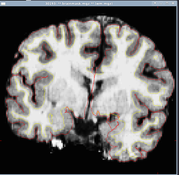
Here's an image (if I attach this correctly) of a particular instance-gray matter excluded in the temporal lobe and pial surface seeming to be below the white matter surface. This seems to be worse on subjects with motion, this one isn't too bad. How would I upload the subject file for you to look at?
Jordan Pierce
Hi Jordan,
it's hard to tell from a single slice but it looks like that might be amygdala, in which case you shouldn't worry about it. In any case, you can upload and we'll take a look
cheers Bruce On Fri, 14 Oct 2011, Jordan Pierce wrote:
Here's an image (if I attach this correctly) of a particular instance-gray matter excluded in the temporal lobe and pial surface seeming to be below the white matter surface. This seems to be worse on subjects with motion, this one isn't too bad. How would I upload the subject file for you to look at?
Jordan Pierce
Yes, it looks like amygdala.
Here are instructions for uploading the case: https://surfer.nmr.mgh.harvard.edu/fswiki/FtpFileExchange
Allison
On Fri, 14 Oct 2011, Bruce Fischl wrote:
Hi Jordan,
it's hard to tell from a single slice but it looks like that might be amygdala, in which case you shouldn't worry about it. In any case, you can upload and we'll take a look
cheers Bruce On Fri, 14 Oct 2011, Jordan Pierce wrote:
Here's an image (if I attach this correctly) of a particular instance-gray matter excluded in the temporal lobe and pial surface seeming to be below the white matter surface. This seems to be worse on subjects with motion, this one isn't too bad. How would I upload the subject file for you to look at?
Jordan Pierce
_______________________________________________ Freesurfer mailing list Freesurfer@nmr.mgh.harvard.edu https://mail.nmr.mgh.harvard.edu/mailman/listinfo/freesurfer
We've uploaded the file as 30193retry.tar.gz
Jordan Pierce
________________________________
Okay, I'll take a look. Allison
On Mon, 17 Oct 2011, Jordan Pierce wrote:
We've uploaded the file as 30193retry.tar.gz Jordan Pierce
_______________________________________________ Freesurfer mailing list Freesurfer@nmr.mgh.harvard.edu https://mail.nmr.mgh.harvard.edu/mailman/listinfo/freesurfer
freesurfer@nmr.mgh.harvard.edu
-
 Allison Player
 -
Allison Stevens
-
 Bruce Fischl
 -
 Jordan Pierce