
Hi everyone!
I'm currently a summer student working at a lab which uses Freesurfer to measure hippocampal volumes. I noticed that scanning the same patient first on a GE 3T (Multi-channel head coil) and then a GE 1.5T (single-channel head coil) 24 hours later resulted in a 30% increase in the left hippocampus volume and a 18% increase for the right hippocampus based on the values in aseg.stats. Can anyone please comment on this and explain the large discrepancies? Thanks!
Jessica Liu

This is not normal. Maybe you should check the aseg volume for improper segmentation.
On Thu, Jun 23, 2011 at 19:27, Jessica Liu jessicaliu92@gmail.com wrote:
Hi everyone!
I'm currently a summer student working at a lab which uses Freesurfer to measure hippocampal volumes. I noticed that scanning the same patient first on a GE 3T (Multi-channel head coil) and then a GE 1.5T (single-channel head coil) 24 hours later resulted in a 30% increase in the left hippocampus volume and a 18% increase for the right hippocampus based on the values in aseg.stats. Can anyone please comment on this and explain the large discrepancies? Thanks!
Jessica Liu
Freesurfer mailing list Freesurfer@nmr.mgh.harvard.edu https://mail.nmr.mgh.harvard.edu/mailman/listinfo/freesurfer
The information in this e-mail is intended only for the person to whom it is addressed. If you believe this e-mail was sent to you in error and the e-mail contains patient information, please contact the Partners Compliance HelpLine at http://www.partners.org/complianceline . If the e-mail was sent to you in error but does not contain patient information, please contact the sender and properly dispose of the e-mail.
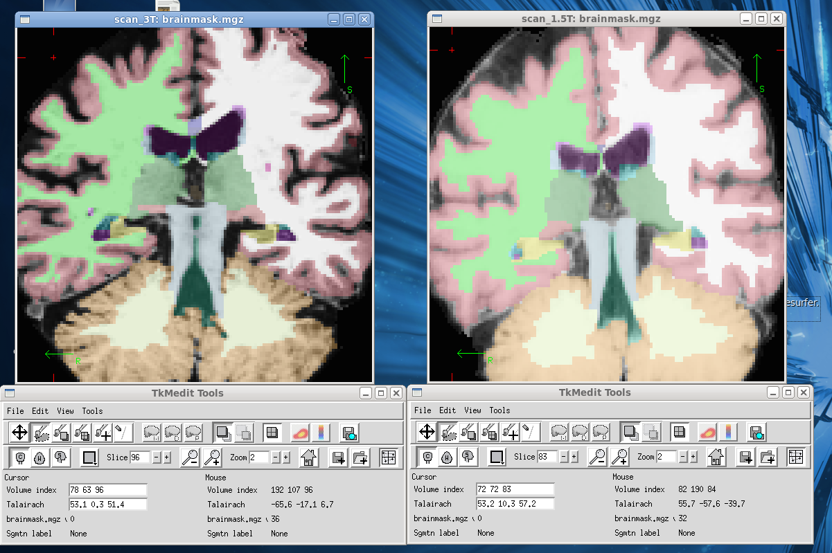
Looking at the scans (attached, 3T on the left and 1.5T on the right, yellow hippocampus), I think you've made a good point. More comments are much appreciated, thanks!
Jessica Liu
2011/6/23 Pedro Paulo de Magalhães Oliveira Junior ppj@netfilter.com.br
This is not normal. Maybe you should check the aseg volume for improper segmentation.
On Thu, Jun 23, 2011 at 19:27, Jessica Liu jessicaliu92@gmail.com wrote:
Hi everyone!
I'm currently a summer student working at a lab which uses Freesurfer to measure hippocampal volumes. I noticed that scanning the same patient first on a GE 3T (Multi-channel head coil) and then a GE 1.5T (single-channel head coil) 24 hours later resulted in a 30% increase in the left hippocampus volume and a 18% increase for the right hippocampus based on the values in aseg.stats. Can anyone please comment on this and explain the large discrepancies? Thanks!
Jessica Liu
Freesurfer mailing list Freesurfer@nmr.mgh.harvard.edu https://mail.nmr.mgh.harvard.edu/mailman/listinfo/freesurfer
The information in this e-mail is intended only for the person to whom it is addressed. If you believe this e-mail was sent to you in error and the e-mail contains patient information, please contact the Partners Compliance HelpLine at http://www.partners.org/complianceline . If the e-mail was sent to you in error but does not contain patient information, please contact the sender and properly dispose of the e-mail.

Hi Jessica,
the 1.5t scan looks kind of washed out and low contrast, but of course it's hard to tell from an image. The 3T looks much crisper. Were the sequence parameters matched? I'm not sure what you're trying to show with this. The 3T multi-channel scan will have a much, much higher SNR than the 1.5T single channel, so the results will not at all be comparable.
Bruce
On Thu, 23 Jun 2011, Jessica Liu wrote:
Looking at the scans (attached, 3T on the left and 1.5T on the right, yellow hippocampus), I think you've made a good point. More comments are much appreciated, thanks!
Jessica Liu
2011/6/23 Pedro Paulo de Magalhães Oliveira Junior ppj@netfilter.com.br This is not normal. Maybe you should check the aseg volume for improper segmentation.
On Thu, Jun 23, 2011 at 19:27, Jessica Liu jessicaliu92@gmail.com wrote: Hi everyone!
I'm currently a summer student working at a lab which uses Freesurfer to measure hippocampal volumes. I noticed that scanning the same patient first on a GE 3T (Multi-channel head coil) and then a GE 1.5T (single-channel head coil) 24 hours later resulted in a 30% increase in the left hippocampus volume and a 18% increase for the right hippocampus based on the values in aseg.stats. Can anyone please comment on this and explain the large discrepancies? Thanks!
Jessica Liu
Freesurfer mailing list Freesurfer@nmr.mgh.harvard.edu https://mail.nmr.mgh.harvard.edu/mailman/listinfo/freesurfer
The information in this e-mail is intended only for the person to whom it is addressed. If you believe this e-mail was sent to you in error and the e-mail contains patient information, please contact the Partners Compliance HelpLine at http://www.partners.org/complianceline . If the e-mail was sent to you in error but does not contain patient information, please contact the sender and properly dispose of the e-mail.
Hi Dr. Fischl,
Thanks for your comments. The aim of my study is to analyze the brain volume in AD subjects. Most of the study was scanned on the 1.5T (single-channel head coil) with a few subjects rescanned on the 3T (12-channel head coil), so we would like to compare the brain volume of the same subject on both scanners. In addition, the protocols on the two scanners didn't match exactly due to the limitations of our over 15-years-old 1.5T scanner (i.e. non-isotropic voxel size). From your comment, I gather that the 1.5T data is rather imprecise/unreliable. Since I'm a summer student, do you recommend that I should concentrate on analyzing the 3T data only, and hopefully all of the subjects can be rescanned on the 3T? Thanks!
Jessica Liu
2011/6/23 Bruce Fischl fischl@nmr.mgh.harvard.edu
Hi Jessica,
the 1.5t scan looks kind of washed out and low contrast, but of course it's hard to tell from an image. The 3T looks much crisper. Were the sequence parameters matched? I'm not sure what you're trying to show with this. The 3T multi-channel scan will have a much, much higher SNR than the 1.5T single channel, so the results will not at all be comparable.
Bruce
On Thu, 23 Jun 2011, Jessica Liu wrote:
Looking at the scans (attached, 3T on the left and 1.5T on the right,
yellow hippocampus), I think you've made a good point. More comments are much appreciated, thanks!
Jessica Liu
2011/6/23 Pedro Paulo de Magalhães Oliveira Junior ppj@netfilter.com.br This is not normal. Maybe you should check the aseg volume for improper segmentation.
On Thu, Jun 23, 2011 at 19:27, Jessica Liu jessicaliu92@gmail.com wrote: Hi everyone!
I'm currently a summer student working at a lab which uses Freesurfer to measure hippocampal volumes. I noticed that scanning the same patient first on a GE 3T (Multi-channel head coil) and then a GE 1.5T (single-channel head coil) 24 hours later resulted in a 30% increase in the left hippocampus volume and a 18% increase for the right hippocampus based on the values in aseg.stats. Can anyone please comment on this and explain the large discrepancies? Thanks!
Jessica Liu
______________________________**_________________ Freesurfer mailing list Freesurfer@nmr.mgh.harvard.edu https://mail.nmr.mgh.harvard.**edu/mailman/listinfo/**freesurferhttps://mail.nmr.mgh.harvard.edu/mailman/listinfo/freesurfer
The information in this e-mail is intended only for the person to whom it is addressed. If you believe this e-mail was sent to you in error and the e-mail contains patient information, please contact the Partners Compliance HelpLine at http://www.partners.org/**compliancelinehttp://www.partners.org/complianceline. If the e-mail was sent to you in error but does not contain patient information, please contact the sender and properly dispose of the e-mail.
Hi Jessica
yes, probably. With so many differences (sequence, field strength, coil) it will be really hard to get anything out of a combined dataset.
sorry Bruce On Fri, 24 Jun 2011, Jessica Liu wrote:
Hi Dr. Fischl,
Thanks for your comments. The aim of my study is to analyze the brain volume in AD subjects. Most of the study was scanned on the 1.5T (single-channel head coil) with a few subjects rescanned on the 3T (12-channel head coil), so we would like to compare the brain volume of the same subject on both scanners. In addition, the protocols on the two scanners didn't match exactly due to the limitations of our over 15-years-old 1.5T scanner (i.e. non-isotropic voxel size). From your comment, I gather that the 1.5T data is rather imprecise/unreliable. Since I'm a summer student, do you recommend that I should concentrate on analyzing the 3T data only, and hopefully all of the subjects can be rescanned on the 3T? Thanks!
Jessica Liu
2011/6/23 Bruce Fischl fischl@nmr.mgh.harvard.edu Hi Jessica,
  the 1.5t scan looks kind of washed out and low contrast, but of
  course it's hard to tell from an image. The 3T looks much
  crisper. Were the sequence parameters matched? I'm not sure what
  you're trying to show with this. The 3T multi-channel scan will
  have a much, much higher SNR than the 1.5T single channel, so
  the results will not at all be comparable.

  Bruce


  On Thu, 23 Jun 2011, Jessica Liu wrote:

  Looking at the scans (attached, 3T on the left and 1.5T on
  the right, yellow
  hippocampus), I think you've made a good point.  More
  comments are much
  appreciated, thanks!

  Jessica Liu

  2011/6/23 Pedro Paulo de Magalhães Oliveira Junior
  <ppj@netfilter.com.br>
       This is not normal. Maybe you should check the aseg
  volume for
       improper segmentation.


  On Thu, Jun 23, 2011 at 19:27, Jessica Liu
  <jessicaliu92@gmail.com>
  wrote:
  Hi everyone!

  I'm currently a summer student working at a lab which uses
  Freesurfer to measure hippocampal volumes.  I noticed that
  scanning the same patient first on a GE 3T (Multi-channel
  head
  coil) and then a GE 1.5T (single-channel head coil) 24
  hours
  later resulted in a 30% increase in the left hippocampus
  volume
  and a 18% increase for the right hippocampus based on the
  values
  in aseg.stats.  Can anyone please comment on this and
  explain
  the large discrepancies?  Thanks!

  Jessica Liu

  _______________________________________________
  Freesurfer mailing list
  Freesurfer@nmr.mgh.harvard.edu
  https://mail.nmr.mgh.harvard.edu/mailman/listinfo/freesurfer


  The information in this e-mail is intended only for the
  person
  to whom it is
  addressed. If you believe this e-mail was sent to you in
  error
  and the e-mail
  contains patient information, please contact the Partners
  Compliance HelpLine at
  http://www.partners.org/complianceline . If the e-mail was
  sent
  to you in error
  but does not contain patient information, please contact
  the
  sender and properly
  dispose of the e-mail.
Alright, thanks for all of your input!
Jessica
2011/6/24 Bruce Fischl fischl@nmr.mgh.harvard.edu
Hi Jessica
yes, probably. With so many differences (sequence, field strength, coil) it will be really hard to get anything out of a combined dataset.
sorry Bruce
On Fri, 24 Jun 2011, Jessica Liu wrote:
Hi Dr. Fischl,
Thanks for your comments. The aim of my study is to analyze the brain volume in AD subjects. Most of the study was scanned on the 1.5T (single-channel head coil) with a few subjects rescanned on the 3T (12-channel head coil), so we would like to compare the brain volume of the same subject on both scanners. In addition, the protocols on the two scanners didn't match exactly due to the limitations of our over 15-years-old 1.5T scanner (i.e. non-isotropic voxel size). From your comment, I gather that the 1.5T data is rather imprecise/unreliable. Since I'm a summer student, do you recommend that I should concentrate on analyzing the 3T data only, and hopefully all of the subjects can be rescanned on the 3T? Thanks!
Jessica Liu
2011/6/23 Bruce Fischl fischl@nmr.mgh.harvard.edu Hi Jessica,
 the 1.5t scan looks kind of washed out and low contrast, but of
 course it's hard to tell from an image. The 3T looks much
 crisper. Were the sequence parameters matched? I'm not sure what
 you're trying to show with this. The 3T multi-channel scan will
 have a much, much higher SNR than the 1.5T single channel, so
 the results will not at all be comparable.

 Bruce


 On Thu, 23 Jun 2011, Jessica Liu wrote:

 Looking at the scans (attached, 3T on the left and 1.5T on
 the right, yellow
 hippocampus), I think you've made a good point.  More
 comments are much
 appreciated, thanks!

 Jessica Liu

 2011/6/23 Pedro Paulo de Magalhães Oliveira Junior
 <ppj@netfilter.com.br>
      This is not normal. Maybe you should check the aseg
 volume for
      improper segmentation.


 On Thu, Jun 23, 2011 at 19:27, Jessica Liu
 <jessicaliu92@gmail.com>
 wrote:
 Hi everyone!

 I'm currently a summer student working at a lab which uses
 Freesurfer to measure hippocampal volumes.  I noticed that
 scanning the same patient first on a GE 3T (Multi-channel
 head
 coil) and then a GE 1.5T (single-channel head coil) 24
 hours
 later resulted in a 30% increase in the left hippocampus
 volume
 and a 18% increase for the right hippocampus based on the
 values
 in aseg.stats.  Can anyone please comment on this and
 explain
 the large discrepancies?  Thanks!

 Jessica Liu

 ______________________________**_________________
 Freesurfer mailing list
 Freesurfer@nmr.mgh.harvard.edu
 https://mail.nmr.mgh.harvard.**edu/mailman/listinfo/**freesurfer<https://mail.nmr.mgh.harvard.edu/mailman/listinfo/freesurfer>


 The information in this e-mail is intended only for the
 person
 to whom it is
 addressed. If you believe this e-mail was sent to you in
 error
 and the e-mail
 contains patient information, please contact the Partners
 Compliance HelpLine at
 http://www.partners.org/**complianceline<http://www.partners.org/complianceline>. If the e-mail was
 sent
 to you in error
 but does not contain patient information, please contact
 the
 sender and properly
 dispose of the e-mail.
Hi Jessica,
what were the coils? Certainly you will find substantial differences comparing across both field strength and receive coil. Did the segmentations look reasonable? Was it the same sequence?
Bruce
On Thu, 23 Jun 2011, Jessica Liu wrote:
Hi everyone!
I'm currently a summer student working at a lab which uses Freesurfer to measure hippocampal volumes. I noticed that scanning the same patient first on a GE 3T (Multi-channel head coil) and then a GE 1.5T (single-channel head coil) 24 hours later resulted in a 30% increase in the left hippocampus volume and a 18% increase for the right hippocampus based on the values in aseg.stats. Can anyone please comment on this and explain the large discrepancies? Thanks!
Jessica Liu
freesurfer@nmr.mgh.harvard.edu
-
 Bruce Fischl
 -
 Jessica Liu
 -
 Pedro Paulo de Magalhães Oliveira Junior