
Hi all,
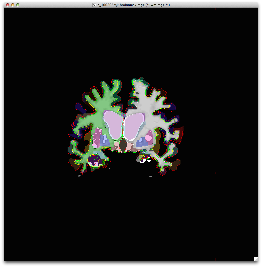
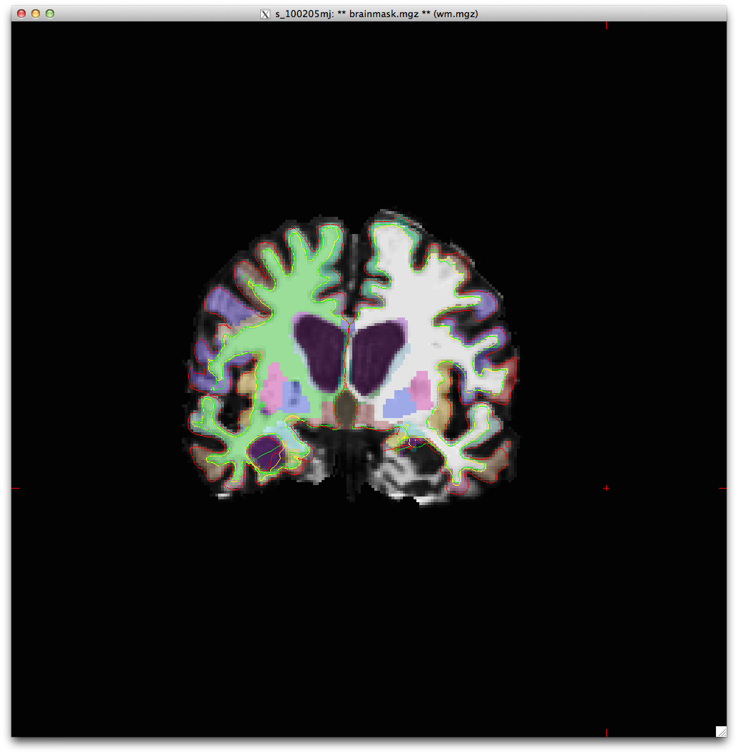
I'm having a bit of trouble with one particular subject for which we brains for 4-5 different time points. The pial surface is consistently misplaced in the same area, see attached main/aux volume images. I'm hesitant to manually replace the white matter myself as I fear it wouldn't be as accurate. Suggestions on other methods to fix this?
The problem is always in the LH temporal right around the lateral inferior ventricle and the fusiform and other subcortical structures.

Hi Jonathan
so you lose that entire gyrus? What are the intensity values of the wm in the brain.mgz in that region? And it's not present in the wm.mgz either? That means it's a segmentation problem not a topology one (which is what I would have guessed)
Bruce On Fri, 28 Jun 2013, Jonathan Holt wrote:
Hi all, I'm having a bit of trouble with one particular subject for which we brains for 4-5 different time points. The pial surface is consistently misplaced in the same area, see attached main/aux volume images. I'm hesitant to manually replace the white matter myself as I fear it wouldn't be as accurate. Suggestions on other methods to fix this?
The problem is always in the LH temporal right around the lateral inferior ventricle and the fusiform and other subcortical structures.
Bruce,
in brainmask.mgz it ranges from about 90-60, as for the wm.mgz values there are only sporadic voxels, most in the 80-90 range. Where the LH inf lat ventricle should be there are some wm voxels at 250.
I agree there are segmentation issues, but there is also a complete lack of WM voxels, what should I do about both?
jon On Jun 28, 2013, at 11:08 AM, Bruce Fischl wrote:
Hi Jonathan
so you lose that entire gyrus? What are the intensity values of the wm in the brain.mgz in that region? And it's not present in the wm.mgz either? That means it's a segmentation problem not a topology one (which is what I would have guessed)
Bruce On Fri, 28 Jun 2013, Jonathan Holt wrote:
Hi all, I'm having a bit of trouble with one particular subject for which we brains for 4-5 different time points. The pial surface is consistently misplaced in the same area, see attached main/aux volume images. I'm hesitant to manually replace the white matter myself as I fear it wouldn't be as accurate. Suggestions on other methods to fix this? The problem is always in the LH temporal right around the lateral inferior ventricle and the fusiform and other subcortical structures.
The information in this e-mail is intended only for the person to whom it is addressed. If you believe this e-mail was sent to you in error and the e-mail contains patient information, please contact the Partners Compliance HelpLine at http://www.partners.org/complianceline . If the e-mail was sent to you in error but does not contain patient information, please contact the sender and properly dispose of the e-mail.
sounds like you'll need control points. Is the WM so dark because of bias fields, or is there some pathology-induced changes to wm intensities?
On Fri, 28 Jun 2013, Jonathan Holt wrote:
Bruce,
in brainmask.mgz it ranges from about 90-60, as for the wm.mgz values there are only sporadic voxels, most in the 80-90 range. Where the LH inf lat ventricle should be there are some wm voxels at 250.
I agree there are segmentation issues, but there is also a complete lack of WM voxels, what should I do about both?
jon On Jun 28, 2013, at 11:08 AM, Bruce Fischl wrote:
Hi Jonathan
so you lose that entire gyrus? What are the intensity values of the wm in the brain.mgz in that region? And it's not present in the wm.mgz either? That means it's a segmentation problem not a topology one (which is what I would have guessed)
Bruce On Fri, 28 Jun 2013, Jonathan Holt wrote:
Hi all, I'm having a bit of trouble with one particular subject for which we brains for 4-5 different time points. The pial surface is consistently misplaced in the same area, see attached main/aux volume images. I'm hesitant to manually replace the white matter myself as I fear it wouldn't be as accurate. Suggestions on other methods to fix this? The problem is always in the LH temporal right around the lateral inferior ventricle and the fusiform and other subcortical structures.
The information in this e-mail is intended only for the person to whom it is addressed. If you believe this e-mail was sent to you in error and the e-mail contains patient information, please contact the Partners Compliance HelpLine at http://www.partners.org/complianceline . If the e-mail was sent to you in error but does not contain patient information, please contact the sender and properly dispose of the e-mail.
Freesurfer mailing list Freesurfer@nmr.mgh.harvard.edu https://mail.nmr.mgh.harvard.edu/mailman/listinfo/freesurfer

Hi Jonathan,
is this in cross sectional processing or in longitudinal?
One thing I notice is significant motion artifacts (ringing), which seem to lead to underestimation of WM, also you could fine tune the skull strip.
Best, Martin
On Jun 28, 2013, at 10:19 AM, Jonathan Holt whatsdac@umich.edu wrote:
Hi all,
I'm having a bit of trouble with one particular subject for which we brains for 4-5 different time points. The pial surface is consistently misplaced in the same area, see attached main/aux volume images. I'm hesitant to manually replace the white matter myself as I fear it wouldn't be as accurate. Suggestions on other methods to fix this?
The problem is always in the LH temporal right around the lateral inferior ventricle and the fusiform and other subcortical structures.
<Screen Shot 2013-06-28 at 10.18.59 AM.png><Screen Shot 2013-06-28 at 10.14.50 AM.png>_______________________________________________ Freesurfer mailing list Freesurfer@nmr.mgh.harvard.edu https://mail.nmr.mgh.harvard.edu/mailman/listinfo/freesurfer
--------------------------------- Dr. Martin Reuter Assistant in Neuroscience - Massachusetts General Hospital Instructor in Neurology - Harvard Medical School MGH / HMS / MIT
A.A.Martinos Center for Biomedical Imaging 149 Thirteenth Street, Suite 2301 Charlestown, MA 02129
Phone: +1-617-724-5652 Email: mreuter@nmr.mgh.harvard.edu reuter@mit.edu Web : http://reuter.mit.edu
Martin,
this is longitudinal processing, would you recommend a more aggressive or less aggressive skull strip?
Bruce,
I've not seen this issue on more than a handful of subjects. They're ALS, but I dont think that is the cause behind this. I'm unfamiliar with bias fields, but it's more likely than pathology.
As for control points, there are no more than 5-7 voxels in that area over the span of 30 slices. Should I simply add to those? On Jun 28, 2013, at 11:19 AM, Martin Reuter wrote:
Hi Jonathan,
is this in cross sectional processing or in longitudinal?
One thing I notice is significant motion artifacts (ringing), which seem to lead to underestimation of WM, also you could fine tune the skull strip.
Best, Martin
On Jun 28, 2013, at 10:19 AM, Jonathan Holt whatsdac@umich.edu wrote:
Hi all,
I'm having a bit of trouble with one particular subject for which we brains for 4-5 different time points. The pial surface is consistently misplaced in the same area, see attached main/aux volume images. I'm hesitant to manually replace the white matter myself as I fear it wouldn't be as accurate. Suggestions on other methods to fix this?
The problem is always in the LH temporal right around the lateral inferior ventricle and the fusiform and other subcortical structures.
<Screen Shot 2013-06-28 at 10.18.59 AM.png><Screen Shot 2013-06-28 at 10.14.50 AM.png>_______________________________________________ Freesurfer mailing list Freesurfer@nmr.mgh.harvard.edu https://mail.nmr.mgh.harvard.edu/mailman/listinfo/freesurfer
Dr. Martin Reuter Assistant in Neuroscience - Massachusetts General Hospital Instructor in Neurology - Harvard Medical School MGH / HMS / MIT
A.A.Martinos Center for Biomedical Imaging 149 Thirteenth Street, Suite 2301 Charlestown, MA 02129
Phone: +1-617-724-5652 Email: mreuter@nmr.mgh.harvard.edu reuter@mit.edu Web : http://reuter.mit.edu
The information in this e-mail is intended only for the person to whom it is addressed. If you believe this e-mail was sent to you in error and the e-mail contains patient information, please contact the Partners Compliance HelpLine at http://www.partners.org/complianceline . If the e-mail was sent to you in error but does not contain patient information, please contact the sender and properly dispose of the e-mail.
oh, if it's that few then yes, but that wasn't what it looked like in the image you sent On Fri, 28 Jun 2013, Jonathan Holt wrote:
Martin, this is longitudinal processing, would you recommend a more aggressive or less aggressive skull strip?
Bruce,
I've not seen this issue on more than a handful of subjects. They're ALS, but I dont think that is the cause behind this. I'm unfamiliar with bias fields, but it's more likely than pathology.
As for control points, there are no more than 5-7 voxels in that area over the span of 30 slices. Should I simply add to those? On Jun 28, 2013, at 11:19 AM, Martin Reuter wrote:
Hi Jonathan,is this in cross sectional processing or in longitudinal?
One thing I notice is significant motion artifacts (ringing), which seem to lead to underestimation of WM, also you could fine tune the skull strip.
Best, Martin
On Jun 28, 2013, at 10:19 AM, Jonathan Holt whatsdac@umich.edu wrote:
Hi all,I'm having a bit of trouble with one particular subject for which we brains for 4-5 different time points. The pial surface is consistently misplaced in the same area, see attached main/aux volume images. I'm hesitant to manually replace the white matter myself as I fear it wouldn't be as accurate. Suggestions on other methods to fix this?
The problem is always in the LH temporal right around the lateral inferior ventricle and the fusiform and other subcortical structures.
<Screen Shot 2013-06-28 at 10.18.59 AM.png><Screen Shot 2013-06-28 at 10.14.50 AM.png>_______________________________________________ Freesurfer mailing list Freesurfer@nmr.mgh.harvard.edu https://mail.nmr.mgh.harvard.edu/mailman/listinfo/freesurfer
Dr. Martin Reuter Assistant in Neuroscience - Massachusetts General Hospital Instructor in Neurology - Harvard Medical School MGH / HMS / MIT
A.A.Martinos Center for Biomedical Imaging 149 Thirteenth Street, Suite 2301 Charlestown, MA 02129
Phone: +1-617-724-5652 Email: mreuter@nmr.mgh.harvard.edu reuter@mit.edu Web : http://reuter.mit.edu
The information in this e-mail is intended only for the person to whom it is addressed. If you believe this e-mail was sent to you in error and the e-mail contains patient information, please contact the Partners Compliance HelpLine at http://www.partners.org/complianceline . If the e-mail was sent to you in error but does not contain patient information, please contact the sender and properly dispose of the e-mail.
Yes, but do these errors show up in the cross sectional data, in the base or in the longitudinal data (or all?)? Does it look good in the base?
I'd suggest more skull strip where the pail grabs dura.
On Jun 28, 2013, at 11:26 AM, Jonathan Holt whatsdac@umich.edu wrote:
Martin,
this is longitudinal processing, would you recommend a more aggressive or less aggressive skull strip?
Bruce,
I've not seen this issue on more than a handful of subjects. They're ALS, but I dont think that is the cause behind this. I'm unfamiliar with bias fields, but it's more likely than pathology.
As for control points, there are no more than 5-7 voxels in that area over the span of 30 slices. Should I simply add to those? On Jun 28, 2013, at 11:19 AM, Martin Reuter wrote:
Hi Jonathan,
is this in cross sectional processing or in longitudinal?
One thing I notice is significant motion artifacts (ringing), which seem to lead to underestimation of WM, also you could fine tune the skull strip.
Best, Martin
On Jun 28, 2013, at 10:19 AM, Jonathan Holt whatsdac@umich.edu wrote:
Hi all,
I'm having a bit of trouble with one particular subject for which we brains for 4-5 different time points. The pial surface is consistently misplaced in the same area, see attached main/aux volume images. I'm hesitant to manually replace the white matter myself as I fear it wouldn't be as accurate. Suggestions on other methods to fix this?
The problem is always in the LH temporal right around the lateral inferior ventricle and the fusiform and other subcortical structures.
<Screen Shot 2013-06-28 at 10.18.59 AM.png><Screen Shot 2013-06-28 at 10.14.50 AM.png>_______________________________________________ Freesurfer mailing list Freesurfer@nmr.mgh.harvard.edu https://mail.nmr.mgh.harvard.edu/mailman/listinfo/freesurfer
Dr. Martin Reuter Assistant in Neuroscience - Massachusetts General Hospital Instructor in Neurology - Harvard Medical School MGH / HMS / MIT
A.A.Martinos Center for Biomedical Imaging 149 Thirteenth Street, Suite 2301 Charlestown, MA 02129
Phone: +1-617-724-5652 Email: mreuter@nmr.mgh.harvard.edu reuter@mit.edu Web : http://reuter.mit.edu
The information in this e-mail is intended only for the person to whom it is addressed. If you believe this e-mail was sent to you in error and the e-mail contains patient information, please contact the Partners Compliance HelpLine at http://www.partners.org/complianceline . If the e-mail was sent to you in error but does not contain patient information, please contact the sender and properly dispose of the e-mail.
--------------------------------- Dr. Martin Reuter Assistant in Neuroscience - Massachusetts General Hospital Instructor in Neurology - Harvard Medical School MGH / HMS / MIT
A.A.Martinos Center for Biomedical Imaging 149 Thirteenth Street, Suite 2301 Charlestown, MA 02129
Phone: +1-617-724-5652 Email: mreuter@nmr.mgh.harvard.edu reuter@mit.edu Web : http://reuter.mit.edu
We have 4 time points for that subject and it's an issue in all 4, to a lesser degree in one of them, but still an issue. Same area, same hemisphere, every time. Perhaps it is pathology based, I am not entirely sure.
On Jun 28, 2013, at 11:19 AM, Martin Reuter wrote:
Hi Jonathan,
is this in cross sectional processing or in longitudinal?
One thing I notice is significant motion artifacts (ringing), which seem to lead to underestimation of WM, also you could fine tune the skull strip.
Best, Martin
On Jun 28, 2013, at 10:19 AM, Jonathan Holt whatsdac@umich.edu wrote:
Hi all,
I'm having a bit of trouble with one particular subject for which we brains for 4-5 different time points. The pial surface is consistently misplaced in the same area, see attached main/aux volume images. I'm hesitant to manually replace the white matter myself as I fear it wouldn't be as accurate. Suggestions on other methods to fix this?
The problem is always in the LH temporal right around the lateral inferior ventricle and the fusiform and other subcortical structures.
<Screen Shot 2013-06-28 at 10.18.59 AM.png><Screen Shot 2013-06-28 at 10.14.50 AM.png>_______________________________________________ Freesurfer mailing list Freesurfer@nmr.mgh.harvard.edu https://mail.nmr.mgh.harvard.edu/mailman/listinfo/freesurfer
Dr. Martin Reuter Assistant in Neuroscience - Massachusetts General Hospital Instructor in Neurology - Harvard Medical School MGH / HMS / MIT
A.A.Martinos Center for Biomedical Imaging 149 Thirteenth Street, Suite 2301 Charlestown, MA 02129
Phone: +1-617-724-5652 Email: mreuter@nmr.mgh.harvard.edu reuter@mit.edu Web : http://reuter.mit.edu
The information in this e-mail is intended only for the person to whom it is addressed. If you believe this e-mail was sent to you in error and the e-mail contains patient information, please contact the Partners Compliance HelpLine at http://www.partners.org/complianceline . If the e-mail was sent to you in error but does not contain patient information, please contact the sender and properly dispose of the e-mail.
So you are not processing this with the longitudinal stream? You are just running each image independently through freesurfer and look at the results? Is there motion in all the 4 time points in this subject or only in the one image that you send? With motion artifacts, surfaces are not very reliable.
Best, martin
On Jun 28, 2013, at 11:36 AM, Jonathan Holt whatsdac@umich.edu wrote:
We have 4 time points for that subject and it's an issue in all 4, to a lesser degree in one of them, but still an issue. Same area, same hemisphere, every time. Perhaps it is pathology based, I am not entirely sure.
On Jun 28, 2013, at 11:19 AM, Martin Reuter wrote:
Hi Jonathan,
is this in cross sectional processing or in longitudinal?
One thing I notice is significant motion artifacts (ringing), which seem to lead to underestimation of WM, also you could fine tune the skull strip.
Best, Martin
On Jun 28, 2013, at 10:19 AM, Jonathan Holt whatsdac@umich.edu wrote:
Hi all,
I'm having a bit of trouble with one particular subject for which we brains for 4-5 different time points. The pial surface is consistently misplaced in the same area, see attached main/aux volume images. I'm hesitant to manually replace the white matter myself as I fear it wouldn't be as accurate. Suggestions on other methods to fix this?
The problem is always in the LH temporal right around the lateral inferior ventricle and the fusiform and other subcortical structures.
<Screen Shot 2013-06-28 at 10.18.59 AM.png><Screen Shot 2013-06-28 at 10.14.50 AM.png>_______________________________________________ Freesurfer mailing list Freesurfer@nmr.mgh.harvard.edu https://mail.nmr.mgh.harvard.edu/mailman/listinfo/freesurfer
Dr. Martin Reuter Assistant in Neuroscience - Massachusetts General Hospital Instructor in Neurology - Harvard Medical School MGH / HMS / MIT
A.A.Martinos Center for Biomedical Imaging 149 Thirteenth Street, Suite 2301 Charlestown, MA 02129
Phone: +1-617-724-5652 Email: mreuter@nmr.mgh.harvard.edu reuter@mit.edu Web : http://reuter.mit.edu
The information in this e-mail is intended only for the person to whom it is addressed. If you believe this e-mail was sent to you in error and the e-mail contains patient information, please contact the Partners Compliance HelpLine at http://www.partners.org/complianceline . If the e-mail was sent to you in error but does not contain patient information, please contact the sender and properly dispose of the e-mail.
--------------------------------- Dr. Martin Reuter Assistant in Neuroscience - Massachusetts General Hospital Instructor in Neurology - Harvard Medical School MGH / HMS / MIT
A.A.Martinos Center for Biomedical Imaging 149 Thirteenth Street, Suite 2301 Charlestown, MA 02129
Phone: +1-617-724-5652 Email: mreuter@nmr.mgh.harvard.edu reuter@mit.edu Web : http://reuter.mit.edu
Martin,
I hate to be noobish, but can you elaborate on the longitudinal stream? It may be that I'm doing entirely too much work.
jon On Jun 28, 2013, at 11:38 AM, Martin Reuter wrote:
So you are not processing this with the longitudinal stream? You are just running each image independently through freesurfer and look at the results? Is there motion in all the 4 time points in this subject or only in the one image that you send? With motion artifacts, surfaces are not very reliable.
Best, martin
On Jun 28, 2013, at 11:36 AM, Jonathan Holt whatsdac@umich.edu wrote:
We have 4 time points for that subject and it's an issue in all 4, to a lesser degree in one of them, but still an issue. Same area, same hemisphere, every time. Perhaps it is pathology based, I am not entirely sure.
On Jun 28, 2013, at 11:19 AM, Martin Reuter wrote:
Hi Jonathan,
is this in cross sectional processing or in longitudinal?
One thing I notice is significant motion artifacts (ringing), which seem to lead to underestimation of WM, also you could fine tune the skull strip.
Best, Martin
On Jun 28, 2013, at 10:19 AM, Jonathan Holt whatsdac@umich.edu wrote:
Hi all,
I'm having a bit of trouble with one particular subject for which we brains for 4-5 different time points. The pial surface is consistently misplaced in the same area, see attached main/aux volume images. I'm hesitant to manually replace the white matter myself as I fear it wouldn't be as accurate. Suggestions on other methods to fix this?
The problem is always in the LH temporal right around the lateral inferior ventricle and the fusiform and other subcortical structures.
<Screen Shot 2013-06-28 at 10.18.59 AM.png><Screen Shot 2013-06-28 at 10.14.50 AM.png>_______________________________________________ Freesurfer mailing list Freesurfer@nmr.mgh.harvard.edu https://mail.nmr.mgh.harvard.edu/mailman/listinfo/freesurfer
Dr. Martin Reuter Assistant in Neuroscience - Massachusetts General Hospital Instructor in Neurology - Harvard Medical School MGH / HMS / MIT
A.A.Martinos Center for Biomedical Imaging 149 Thirteenth Street, Suite 2301 Charlestown, MA 02129
Phone: +1-617-724-5652 Email: mreuter@nmr.mgh.harvard.edu reuter@mit.edu Web : http://reuter.mit.edu
The information in this e-mail is intended only for the person to whom it is addressed. If you believe this e-mail was sent to you in error and the e-mail contains patient information, please contact the Partners Compliance HelpLine at http://www.partners.org/complianceline . If the e-mail was sent to you in error but does not contain patient information, please contact the sender and properly dispose of the e-mail.
Dr. Martin Reuter Assistant in Neuroscience - Massachusetts General Hospital Instructor in Neurology - Harvard Medical School MGH / HMS / MIT
A.A.Martinos Center for Biomedical Imaging 149 Thirteenth Street, Suite 2301 Charlestown, MA 02129
Phone: +1-617-724-5652 Email: mreuter@nmr.mgh.harvard.edu reuter@mit.edu Web : http://reuter.mit.edu
Hi Jon,
The longitudinal stream in freesurfer is designed to reduce variability in situations where you have several scans from the same subject. The information, that it is the same subject, is valuable and by initializing many steps in recon-all with common data we can increase reliability of measures. The stream has three steps 1. process your data independently (called cross) 2. create a within-subject template and process that (called base) 3. initialize each time point with information from the template (called long).
you can find some more info here: https://surfer.nmr.mgh.harvard.edu/fswiki/LongitudinalProcessing
and here: Within-Subject Template Estimation for Unbiased Longitudinal Image Analysis M. Reuter, N.J. Schmansky, H.D. Rosas, B. Fischl. NeuroImage 61(4), pp. 1402-1418, 2012. http://reuter.mit.edu/papers/reuter-long12.pdf
Not sure if this will fix things for you. If the image quality is bad and if the cross sectional stream (first step) has severe problems already, it is unlikely that everything will recover without any edits. But if, for example, one time point has motion artifiacts and the others not, then the template will be fine and there is a good chance that results on all time points will be good after the 3rd step.
Best, Martin
On Jun 28, 2013, at 9:14 PM, Jonathan Holt whatsdac@umich.edu wrote:
Martin,
I hate to be noobish, but can you elaborate on the longitudinal stream? It may be that I'm doing entirely too much work.
jon On Jun 28, 2013, at 11:38 AM, Martin Reuter wrote:
So you are not processing this with the longitudinal stream? You are just running each image independently through freesurfer and look at the results? Is there motion in all the 4 time points in this subject or only in the one image that you send? With motion artifacts, surfaces are not very reliable.
Best, martin
On Jun 28, 2013, at 11:36 AM, Jonathan Holt whatsdac@umich.edu wrote:
We have 4 time points for that subject and it's an issue in all 4, to a lesser degree in one of them, but still an issue. Same area, same hemisphere, every time. Perhaps it is pathology based, I am not entirely sure.
On Jun 28, 2013, at 11:19 AM, Martin Reuter wrote:
Hi Jonathan,
is this in cross sectional processing or in longitudinal?
One thing I notice is significant motion artifacts (ringing), which seem to lead to underestimation of WM, also you could fine tune the skull strip.
Best, Martin
On Jun 28, 2013, at 10:19 AM, Jonathan Holt whatsdac@umich.edu wrote:
Hi all,
I'm having a bit of trouble with one particular subject for which we brains for 4-5 different time points. The pial surface is consistently misplaced in the same area, see attached main/aux volume images. I'm hesitant to manually replace the white matter myself as I fear it wouldn't be as accurate. Suggestions on other methods to fix this?
The problem is always in the LH temporal right around the lateral inferior ventricle and the fusiform and other subcortical structures.
<Screen Shot 2013-06-28 at 10.18.59 AM.png><Screen Shot 2013-06-28 at 10.14.50 AM.png>_______________________________________________ Freesurfer mailing list Freesurfer@nmr.mgh.harvard.edu https://mail.nmr.mgh.harvard.edu/mailman/listinfo/freesurfer
Dr. Martin Reuter Assistant in Neuroscience - Massachusetts General Hospital Instructor in Neurology - Harvard Medical School MGH / HMS / MIT
A.A.Martinos Center for Biomedical Imaging 149 Thirteenth Street, Suite 2301 Charlestown, MA 02129
Phone: +1-617-724-5652 Email: mreuter@nmr.mgh.harvard.edu reuter@mit.edu Web : http://reuter.mit.edu
The information in this e-mail is intended only for the person to whom it is addressed. If you believe this e-mail was sent to you in error and the e-mail contains patient information, please contact the Partners Compliance HelpLine at http://www.partners.org/complianceline . If the e-mail was sent to you in error but does not contain patient information, please contact the sender and properly dispose of the e-mail.
Dr. Martin Reuter Assistant in Neuroscience - Massachusetts General Hospital Instructor in Neurology - Harvard Medical School MGH / HMS / MIT
A.A.Martinos Center for Biomedical Imaging 149 Thirteenth Street, Suite 2301 Charlestown, MA 02129
Phone: +1-617-724-5652 Email: mreuter@nmr.mgh.harvard.edu reuter@mit.edu Web : http://reuter.mit.edu
--------------------------------- Dr. Martin Reuter Assistant in Neuroscience - Massachusetts General Hospital Instructor in Neurology - Harvard Medical School MGH / HMS / MIT
A.A.Martinos Center for Biomedical Imaging 149 Thirteenth Street, Suite 2301 Charlestown, MA 02129
Phone: +1-617-724-5652 Email: mreuter@nmr.mgh.harvard.edu reuter@mit.edu Web : http://reuter.mit.edu
Martin,
I'm going to experiment with data with better image quality using the longitudinal stream. In the first step of the workflow I'm confused to what 'dcm' stands for in the following command
recon-all -all -s <tpNid> -i path_to_tpN_dcm
could you elaborate?
jon
On Sat, Jun 29, 2013 at 12:29 AM, Martin Reuter <mreuter@nmr.mgh.harvard.edu
wrote:
Hi Jon,
The longitudinal stream in freesurfer is designed to reduce variability in situations where you have several scans from the same subject. The information, that it is the same subject, is valuable and by initializing many steps in recon-all with common data we can increase reliability of measures. The stream has three steps
- process your data independently (called cross)
- create a within-subject template and process that (called base)
- initialize each time point with information from the template (called
long).
you can find some more info here: https://surfer.nmr.mgh.harvard.edu/fswiki/LongitudinalProcessing
and here: Within-Subject Template Estimation for Unbiased Longitudinal Image Analysis M. Reuter, N.J. Schmansky, H.D. Rosas, B. Fischl. NeuroImage 61(4), pp. 1402-1418, 2012. http://reuter.mit.edu/papers/reuter-long12.pdf
Not sure if this will fix things for you. If the image quality is bad and if the cross sectional stream (first step) has severe problems already, it is unlikely that everything will recover without any edits. But if, for example, one time point has motion artifiacts and the others not, then the template will be fine and there is a good chance that results on all time points will be good after the 3rd step.
Best, Martin
On Jun 28, 2013, at 9:14 PM, Jonathan Holt whatsdac@umich.edu wrote:
Martin,
I hate to be noobish, but can you elaborate on the longitudinal stream? It may be that I'm doing entirely too much work.
jon On Jun 28, 2013, at 11:38 AM, Martin Reuter wrote:
So you are not processing this with the longitudinal stream? You are just running each image independently through freesurfer and look at the results? Is there motion in all the 4 time points in this subject or only in the one image that you send? With motion artifacts, surfaces are not very reliable.
Best, martin
On Jun 28, 2013, at 11:36 AM, Jonathan Holt whatsdac@umich.edu wrote:
We have 4 time points for that subject and it's an issue in all 4, to a lesser degree in one of them, but still an issue. Same area, same hemisphere, every time. Perhaps it is pathology based, I am not entirely sure.
On Jun 28, 2013, at 11:19 AM, Martin Reuter wrote:
Hi Jonathan,
is this in cross sectional processing or in longitudinal?
One thing I notice is significant motion artifacts (ringing), which seem to lead to underestimation of WM, also you could fine tune the skull strip.
Best, Martin
On Jun 28, 2013, at 10:19 AM, Jonathan Holt whatsdac@umich.edu wrote:
Hi all,
I'm having a bit of trouble with one particular subject for which we brains for 4-5 different time points. The pial surface is consistently misplaced in the same area, see attached main/aux volume images. I'm hesitant to manually replace the white matter myself as I fear it wouldn't be as accurate. Suggestions on other methods to fix this?
The problem is always in the LH temporal right around the lateral inferior ventricle and the fusiform and other subcortical structures.
<Screen Shot 2013-06-28 at 10.18.59 AM.png><Screen Shot 2013-06-28 at 10.14.50 AM.png>_______________________________________________ Freesurfer mailing list Freesurfer@nmr.mgh.harvard.edu https://mail.nmr.mgh.harvard.edu/mailman/listinfo/freesurfer
Dr. Martin Reuter Assistant in Neuroscience - Massachusetts General Hospital Instructor in Neurology - Harvard Medical School MGH / HMS / MIT
A.A.Martinos Center for Biomedical Imaging 149 Thirteenth Street, Suite 2301 Charlestown, MA 02129
Phone: +1-617-724-5652 Email: mreuter@nmr.mgh.harvard.edu reuter@mit.edu Web : http://reuter.mit.edu
The information in this e-mail is intended only for the person to whom it is addressed. If you believe this e-mail was sent to you in error and the e-mail contains patient information, please contact the Partners Compliance HelpLine at http://www.partners.org/complianceline . If the e-mail was sent to you in error but does not contain patient information, please contact the sender and properly dispose of the e-mail.
Dr. Martin Reuter Assistant in Neuroscience - Massachusetts General Hospital Instructor in Neurology - Harvard Medical School MGH / HMS / MIT
A.A.Martinos Center for Biomedical Imaging 149 Thirteenth Street, Suite 2301 Charlestown, MA 02129
Phone: +1-617-724-5652 Email: mreuter@nmr.mgh.harvard.edu reuter@mit.edu Web : http://reuter.mit.edu
Hi Jon
That is one way of calling the regular recon-all stream. You point it to any input file with the -i flag. Here dcm could be a dicom image, but can be nifti or another format.
Best, Martin
On 07/01/2013 10:17 AM, Jonathan Holt wrote:
Martin,
I'm going to experiment with data with better image quality using the longitudinal stream. In the first step of the workflow I'm confused to what 'dcm' stands for in the following command
recon-all -all -s <tpNid> -i path_to_tpN_dcm
could you elaborate?
jon
On Sat, Jun 29, 2013 at 12:29 AM, Martin Reuter <mreuter@nmr.mgh.harvard.edu mailto:mreuter@nmr.mgh.harvard.edu> wrote:
Hi Jon, The longitudinal stream in freesurfer is designed to reduce variability in situations where you have several scans from the same subject. The information, that it is the same subject, is valuable and by initializing many steps in recon-all with common data we can increase reliability of measures. The stream has three steps 1. process your data independently (called cross) 2. create a within-subject template and process that (called base) 3. initialize each time point with information from the template (called long). you can find some more info here: https://surfer.nmr.mgh.harvard.edu/fswiki/LongitudinalProcessing and here: Within-Subject Template Estimation for Unbiased Longitudinal Image Analysis M. Reuter, N.J. Schmansky, H.D. Rosas, B. Fischl. NeuroImage 61(4), pp. 1402-1418, 2012. http://reuter.mit.edu/papers/reuter-long12.pdf Not sure if this will fix things for you. If the image quality is bad and if the cross sectional stream (first step) has severe problems already, it is unlikely that everything will recover without any edits. But if, for example, one time point has motion artifiacts and the others not, then the template will be fine and there is a good chance that results on all time points will be good after the 3rd step. Best, Martin On Jun 28, 2013, at 9:14 PM, Jonathan Holt <whatsdac@umich.edu <mailto:whatsdac@umich.edu>> wrote:Martin, I hate to be noobish, but can you elaborate on the longitudinal stream? It may be that I'm doing entirely too much work. jon On Jun 28, 2013, at 11:38 AM, Martin Reuter wrote:So you are not processing this with the longitudinal stream? You are just running each image independently through freesurfer and look at the results? Is there motion in all the 4 time points in this subject or only in the one image that you send? With motion artifacts, surfaces are not very reliable. Best, martin On Jun 28, 2013, at 11:36 AM, Jonathan Holt <whatsdac@umich.edu <mailto:whatsdac@umich.edu>> wrote:We have 4 time points for that subject and it's an issue in all 4, to a lesser degree in one of them, but still an issue. Same area, same hemisphere, every time. Perhaps it is pathology based, I am not entirely sure. On Jun 28, 2013, at 11:19 AM, Martin Reuter wrote:Hi Jonathan, is this in cross sectional processing or in longitudinal? One thing I notice is significant motion artifacts (ringing), which seem to lead to underestimation of WM, also you could fine tune the skull strip. Best, Martin On Jun 28, 2013, at 10:19 AM, Jonathan Holt <whatsdac@umich.edu <mailto:whatsdac@umich.edu>> wrote:Hi all, I'm having a bit of trouble with one particular subject for which we brains for 4-5 different time points. The pial surface is consistently misplaced in the same area, see attached main/aux volume images. I'm hesitant to manually replace the white matter myself as I fear it wouldn't be as accurate. Suggestions on other methods to fix this? The problem is always in the LH temporal right around the lateral inferior ventricle and the fusiform and other subcortical structures. <Screen Shot 2013-06-28 at 10.18.59 AM.png><Screen Shot 2013-06-28 at 10.14.50 AM.png>_______________________________________________ Freesurfer mailing list Freesurfer@nmr.mgh.harvard.edu <mailto:Freesurfer@nmr.mgh.harvard.edu> https://mail.nmr.mgh.harvard.edu/mailman/listinfo/freesurfer--------------------------------- Dr. Martin Reuter Assistant in Neuroscience - Massachusetts General Hospital Instructor in Neurology - Harvard Medical School MGH / HMS / MIT A.A.Martinos Center for Biomedical Imaging 149 Thirteenth Street, Suite 2301 Charlestown, MA 02129 Phone: +1-617-724-5652 <tel:%2B1-617-724-5652> Email: mreuter@nmr.mgh.harvard.edu <mailto:mreuter@nmr.mgh.harvard.edu> reuter@mit.edu <mailto:reuter@mit.edu> Web : http://reuter.mit.edu <http://reuter.mit.edu/> The information in this e-mail is intended only for the person to whom it is addressed. If you believe this e-mail was sent to you in error and the e-mail contains patient information, please contact the Partners Compliance HelpLine at http://www.partners.org/complianceline . If the e-mail was sent to you in error but does not contain patient information, please contact the sender and properly dispose of the e-mail.--------------------------------- Dr. Martin Reuter Assistant in Neuroscience - Massachusetts General Hospital Instructor in Neurology - Harvard Medical School MGH / HMS / MIT A.A.Martinos Center for Biomedical Imaging 149 Thirteenth Street, Suite 2301 Charlestown, MA 02129 Phone: +1-617-724-5652 <tel:%2B1-617-724-5652> Email: mreuter@nmr.mgh.harvard.edu <mailto:mreuter@nmr.mgh.harvard.edu> reuter@mit.edu <mailto:reuter@mit.edu> Web : http://reuter.mit.edu <http://reuter.mit.edu/>--------------------------------- Dr. Martin Reuter Assistant in Neuroscience - Massachusetts General Hospital Instructor in Neurology - Harvard Medical School MGH / HMS / MIT A.A.Martinos Center for Biomedical Imaging 149 Thirteenth Street, Suite 2301 Charlestown, MA 02129 Phone: +1-617-724-5652 <tel:%2B1-617-724-5652> Email: mreuter@nmr.mgh.harvard.edu <mailto:mreuter@nmr.mgh.harvard.edu> reuter@mit.edu <mailto:reuter@mit.edu> Web : http://reuter.mit.edu <http://reuter.mit.edu/>
OK,
so I should plug the nifti's as opposed to the freesurfer generated mgzs
jon On Jul 1, 2013, at 11:35 AM, Martin Reuter wrote:
Hi Jon
That is one way of calling the regular recon-all stream. You point it to any input file with the -i flag. Here dcm could be a dicom image, but can be nifti or another format.
Best, Martin
On 07/01/2013 10:17 AM, Jonathan Holt wrote:
Martin,
I'm going to experiment with data with better image quality using the longitudinal stream. In the first step of the workflow I'm confused to what 'dcm' stands for in the following command
recon-all -all -s <tpNid> -i path_to_tpN_dcm
could you elaborate?
jon
On Sat, Jun 29, 2013 at 12:29 AM, Martin Reuter mreuter@nmr.mgh.harvard.edu wrote: Hi Jon,
The longitudinal stream in freesurfer is designed to reduce variability in situations where you have several scans from the same subject. The information, that it is the same subject, is valuable and by initializing many steps in recon-all with common data we can increase reliability of measures. The stream has three steps
- process your data independently (called cross)
- create a within-subject template and process that (called base)
- initialize each time point with information from the template (called long).
you can find some more info here: https://surfer.nmr.mgh.harvard.edu/fswiki/LongitudinalProcessing
and here: Within-Subject Template Estimation for Unbiased Longitudinal Image Analysis M. Reuter, N.J. Schmansky, H.D. Rosas, B. Fischl. NeuroImage 61(4), pp. 1402-1418, 2012. http://reuter.mit.edu/papers/reuter-long12.pdf
Not sure if this will fix things for you. If the image quality is bad and if the cross sectional stream (first step) has severe problems already, it is unlikely that everything will recover without any edits. But if, for example, one time point has motion artifiacts and the others not, then the template will be fine and there is a good chance that results on all time points will be good after the 3rd step.
Best, Martin
On Jun 28, 2013, at 9:14 PM, Jonathan Holt whatsdac@umich.edu wrote:
Martin,

I hate to be noobish, but can you elaborate on the longitudinal
stream? It may be that I'm doing entirely too much work.

jon
On Jun 28, 2013, at 11:38 AM, Martin Reuter wrote:
So you are not processing this with the longitudinal stream? You are just running each image independently through freesurfer and look at the results? Is there motion in all the 4 time points in this subject or only in the one image that you send? With motion artifacts, surfaces are not very reliable.
Best, martin
On Jun 28, 2013, at 11:36 AM, Jonathan Holt whatsdac@umich.edu wrote:
We have 4 time points for that subject and it's an issue in all
4, to a lesser degree in one of them, but still an issue. Same
area, same hemisphere, every time. Perhaps it is pathology
based, I am not entirely sure.


On Jun 28, 2013, at 11:19 AM, Martin Reuter wrote:
Hi Jonathan,
is this in cross sectional processing or in longitudinal?
One thing I notice is significant motion artifacts (ringing), which seem to lead to underestimation of WM, also you could fine tune the skull strip.
Best, Martin
On Jun 28, 2013, at 10:19 AM, Jonathan Holt whatsdac@umich.edu wrote:
> Hi all, > > I'm having a bit of trouble with one particular subject for which we brains for 4-5 different time points. The pial surface is consistently misplaced in the same area, see attached main/aux volume images. I'm hesitant to manually replace the white matter myself as I fear it wouldn't be as accurate. Suggestions on other methods to fix this? > > The problem is always in the LH temporal right around the lateral inferior ventricle and the fusiform and other subcortical structures. > > > <Screen Shot 2013-06-28 at 10.18.59 AM.png><Screen Shot 2013-06-28 at 10.14.50 AM.png>_______________________________________________ > Freesurfer mailing list > Freesurfer@nmr.mgh.harvard.edu > https://mail.nmr.mgh.harvard.edu/mailman/listinfo/freesurfer
Dr. Martin Reuter Assistant in Neuroscience - Massachusetts General Hospital Instructor in Neurology - Harvard Medical School MGH / HMS / MIT
A.A.Martinos Center for Biomedical Imaging 149 Thirteenth Street, Suite 2301 Charlestown, MA 02129
Phone: +1-617-724-5652 Email: mreuter@nmr.mgh.harvard.edu reuter@mit.edu Web : http://reuter.mit.edu
The information in this e-mail is intended only for the person to whom it is addressed. If you believe this e-mail was sent to you in error and the e-mail contains patient information, please contact the Partners Compliance HelpLine at http://www.partners.org/complianceline . If the e-mail was sent to you in error but does not contain patient information, please contact the sender and properly dispose of the e-mail.
Dr. Martin Reuter Assistant in Neuroscience - Massachusetts General Hospital Instructor in Neurology - Harvard Medical School MGH / HMS / MIT
A.A.Martinos Center for Biomedical Imaging 149 Thirteenth Street, Suite 2301 Charlestown, MA 02129
Phone: +1-617-724-5652 Email: mreuter@nmr.mgh.harvard.edu reuter@mit.edu Web : http://reuter.mit.edu
Dr. Martin Reuter Assistant in Neuroscience - Massachusetts General Hospital Instructor in Neurology - Harvard Medical School MGH / HMS / MIT
A.A.Martinos Center for Biomedical Imaging 149 Thirteenth Street, Suite 2301 Charlestown, MA 02129
Phone: +1-617-724-5652 Email: mreuter@nmr.mgh.harvard.edu reuter@mit.edu Web : http://reuter.mit.edu
-- Martin Reuter, Ph.D. Assistant in Neuroscience - Massachusetts General Hospital Instructor in Neurology - Harvard Medical School MGH / HMS / MIT
A.A.Martinos Center for Biomedical Imaging 149 Thirteenth Street, Suite 2301 Charlestown, MA 02129
Phone: +1-617-724-5652 Email: mreuter@nmr.mgh.harvard.edu reuter@mit.edu Web : http://reuter.mit.edu
Hi Jon,
this is for the cross sectional stream, so it is the same as what you did when you processed your data independently. You point recon all to some input file and it generates the subject/mri/orig/001.mgz etc . You can also do this in two steps, the -i to import your data and then the -all to run everything. Or you can manually create subject/mri/orig/ and place the 001.mgz file there if you like and then run -all and no -i.
Best, Martin
On 07/01/2013 12:28 PM, Jonathan Holt wrote:
OK,
so I should plug the nifti's as opposed to the freesurfer generated mgzs
jon On Jul 1, 2013, at 11:35 AM, Martin Reuter wrote:
Hi Jon
That is one way of calling the regular recon-all stream. You point it to any input file with the -i flag. Here dcm could be a dicom image, but can be nifti or another format.
Best, Martin
On 07/01/2013 10:17 AM, Jonathan Holt wrote:
Martin,
I'm going to experiment with data with better image quality using the longitudinal stream. In the first step of the workflow I'm confused to what 'dcm' stands for in the following command
recon-all -all -s <tpNid> -i path_to_tpN_dcm
could you elaborate?
jon
On Sat, Jun 29, 2013 at 12:29 AM, Martin Reuter <mreuter@nmr.mgh.harvard.edu mailto:mreuter@nmr.mgh.harvard.edu> wrote:
Hi Jon, The longitudinal stream in freesurfer is designed to reduce variability in situations where you have several scans from the same subject. The information, that it is the same subject, is valuable and by initializing many steps in recon-all with common data we can increase reliability of measures. The stream has three steps 1. process your data independently (called cross) 2. create a within-subject template and process that (called base) 3. initialize each time point with information from the template (called long). you can find some more info here: https://surfer.nmr.mgh.harvard.edu/fswiki/LongitudinalProcessing and here: Within-Subject Template Estimation for Unbiased Longitudinal Image Analysis M. Reuter, N.J. Schmansky, H.D. Rosas, B. Fischl. NeuroImage 61(4), pp. 1402-1418, 2012. http://reuter.mit.edu/papers/reuter-long12.pdf Not sure if this will fix things for you. If the image quality is bad and if the cross sectional stream (first step) has severe problems already, it is unlikely that everything will recover without any edits. But if, for example, one time point has motion artifiacts and the others not, then the template will be fine and there is a good chance that results on all time points will be good after the 3rd step. Best, Martin On Jun 28, 2013, at 9:14 PM, Jonathan Holt <whatsdac@umich.edu <mailto:whatsdac@umich.edu>> wrote:Martin, I hate to be noobish, but can you elaborate on the longitudinal stream? It may be that I'm doing entirely too much work. jon On Jun 28, 2013, at 11:38 AM, Martin Reuter wrote:So you are not processing this with the longitudinal stream? You are just running each image independently through freesurfer and look at the results? Is there motion in all the 4 time points in this subject or only in the one image that you send? With motion artifacts, surfaces are not very reliable. Best, martin On Jun 28, 2013, at 11:36 AM, Jonathan Holt <whatsdac@umich.edu <mailto:whatsdac@umich.edu>> wrote:We have 4 time points for that subject and it's an issue in all 4, to a lesser degree in one of them, but still an issue. Same area, same hemisphere, every time. Perhaps it is pathology based, I am not entirely sure. On Jun 28, 2013, at 11:19 AM, Martin Reuter wrote:> Hi Jonathan, > > is this in cross sectional processing or in longitudinal? > > One thing I notice is significant motion artifacts > (ringing), which seem to lead to underestimation of WM, also > you could fine tune the skull strip. > > Best, Martin > > On Jun 28, 2013, at 10:19 AM, Jonathan Holt > <whatsdac@umich.edu mailto:whatsdac@umich.edu> wrote: > >> Hi all, >> >> I'm having a bit of trouble with one particular subject for >> which we brains for 4-5 different time points. The pial >> surface is consistently misplaced in the same area, see >> attached main/aux volume images. I'm hesitant to manually >> replace the white matter myself as I fear it wouldn't be as >> accurate. Suggestions on other methods to fix this? >> >> The problem is always in the LH temporal right around the >> lateral inferior ventricle and the fusiform and other >> subcortical structures. >> >> >> <Screen Shot 2013-06-28 at 10.18.59 AM.png><Screen Shot >> 2013-06-28 at 10.14.50 >> AM.png>_______________________________________________ >> Freesurfer mailing list >> Freesurfer@nmr.mgh.harvard.edu >> mailto:Freesurfer@nmr.mgh.harvard.edu >> https://mail.nmr.mgh.harvard.edu/mailman/listinfo/freesurfer > > --------------------------------- > Dr. Martin Reuter > Assistant in Neuroscience - Massachusetts General Hospital > Instructor in Neurology - Harvard Medical School > MGH / HMS / MIT > > A.A.Martinos Center for Biomedical Imaging > 149 Thirteenth Street, Suite 2301 > Charlestown, MA 02129 > > Phone: +1-617-724-5652 tel:%2B1-617-724-5652 > Email: > mreuter@nmr.mgh.harvard.edu mailto:mreuter@nmr.mgh.harvard.edu > reuter@mit.edu mailto:reuter@mit.edu > Web : http://reuter.mit.edu http://reuter.mit.edu/ > > > The information in this e-mail is intended only for the > person to whom it is > addressed. If you believe this e-mail was sent to you in > error and the e-mail > contains patient information, please contact the Partners > Compliance HelpLine at > http://www.partners.org/complianceline . If the e-mail was > sent to you in error > but does not contain patient information, please contact the > sender and properly > dispose of the e-mail. >
---------------------------------
Dr. Martin Reuter
Assistant in Neuroscience - Massachusetts General Hospital
Instructor in Neurology   - Harvard Medical School
MGH / HMS / MIT

A.A.Martinos Center for Biomedical Imaging
149 Thirteenth Street, Suite 2301
Charlestown, MA 02129

Phone: +1-617-724-5652 <tel:%2B1-617-724-5652>
Email:
mreuter@nmr.mgh.harvard.edu <mailto:mreuter@nmr.mgh.harvard.edu>
reuter@mit.edu <mailto:reuter@mit.edu>
Web  : http://reuter.mit.edu <http://reuter.mit.edu/>
--------------------------------- Dr. Martin Reuter Assistant in Neuroscience - Massachusetts General Hospital Instructor in Neurology - Harvard Medical School MGH / HMS / MIT A.A.Martinos Center for Biomedical Imaging 149 Thirteenth Street, Suite 2301 Charlestown, MA 02129 Phone: +1-617-724-5652 <tel:%2B1-617-724-5652> Email: mreuter@nmr.mgh.harvard.edu <mailto:mreuter@nmr.mgh.harvard.edu> reuter@mit.edu <mailto:reuter@mit.edu> Web : http://reuter.mit.edu <http://reuter.mit.edu/>-- Martin Reuter, Ph.D. Assistant in Neuroscience - Massachusetts General Hospital Instructor in Neurology - Harvard Medical School MGH / HMS / MIT
A.A.Martinos Center for Biomedical Imaging 149 Thirteenth Street, Suite 2301 Charlestown, MA 02129
Phone: +1-617-724-5652 Email: mreuter@nmr.mgh.harvard.edu reuter@mit.edu Web :http://reuter.mit.edu
my guess is it that it is, as for whatever reason temporal WM seems to darken first in an array of disorders. I would start with a couple of control points
cheers Bruce
On Fri, 28 Jun 2013, Jonathan Holt wrote:
We have 4 time points for that subject and it's an issue in
all 4, to a lesser degree in one of them, but still an issue.
Same area, same hemisphere, every time. Perhaps it is
pathology based, I am not entirely sure.


On Jun 28, 2013, at 11:19 AM, Martin Reuter wrote:
Hi Jonathan,is this in cross sectional processing or in longitudinal?
One thing I notice is significant motion artifacts (ringing), which seem to lead to underestimation of WM, also you could fine tune the skull strip.
Best, Martin
On Jun 28, 2013, at 10:19 AM, Jonathan Holt whatsdac@umich.edu wrote:
Hi all,I'm having a bit of trouble with one particular subject for which we brains for 4-5 different time points. The pial surface is consistently misplaced in the same area, see attached main/aux volume images. I'm hesitant to manually replace the white matter myself as I fear it wouldn't be as accurate. Suggestions on other methods to fix this?
The problem is always in the LH temporal right around the lateral inferior ventricle and the fusiform and other subcortical structures.
<Screen Shot 2013-06-28 at 10.18.59 AM.png><Screen Shot 2013-06-28 at 10.14.50 AM.png>_______________________________________________ Freesurfer mailing list Freesurfer@nmr.mgh.harvard.edu https://mail.nmr.mgh.harvard.edu/mailman/listinfo/freesurfer
Dr. Martin Reuter Assistant in Neuroscience - Massachusetts General Hospital Instructor in Neurology - Harvard Medical School MGH / HMS / MIT
A.A.Martinos Center for Biomedical Imaging 149 Thirteenth Street, Suite 2301 Charlestown, MA 02129
Phone: +1-617-724-5652 Email: mreuter@nmr.mgh.harvard.edu reuter@mit.edu Web : http://reuter.mit.edu
The information in this e-mail is intended only for the person to whom it is addressed. If you believe this e-mail was sent to you in error and the e-mail contains patient information, please contact the Partners Compliance HelpLine at http://www.partners.org/complianceline . If the e-mail was sent to you in error but does not contain patient information, please contact the sender and properly dispose of the e-mail.
freesurfer@nmr.mgh.harvard.edu
-
 Bruce Fischl
 -
 Jonathan Holt
 -
 Martin Reuter