
Dear Experts,
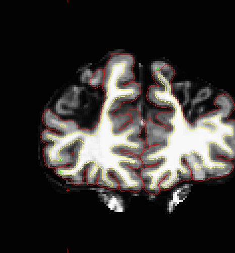
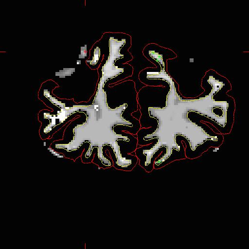
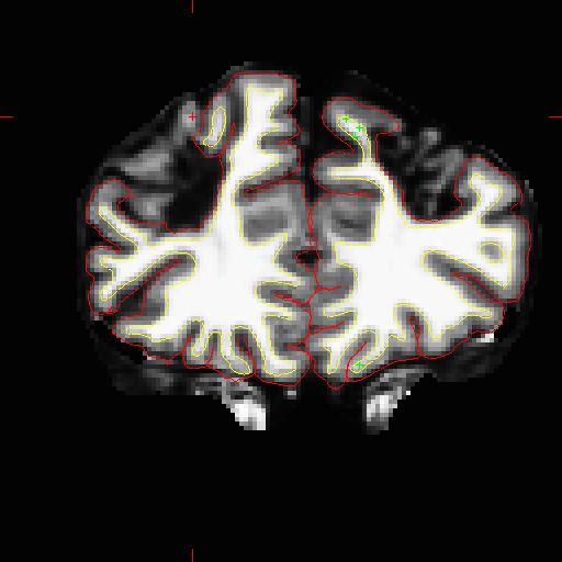
I have a subject with severe atrophy in bilateral frontal and parietal regions and after many rounds of edits to the brainmask, wm.mgz, and control points, these regions are still left uncaptured. Is there a way to edit the pial surface to include these grey matter regions? I am hesitant to draw them in via the wm surface since there is no definite WM present.
I do have a FLAIR also on this subject.
I've attached a couple examples.
Thanks!
Corinna

Sorry Corinna
I meant to answer this the first time but it slipped through the cracks. I doubt there is much you can do if there is that big a lesion. If the topology is wrong and you can't see tissue in those regions it's going to be very hard Bruce
On Wed, 10 Feb 2016, Corinna Bauer wrote:
Dear Experts,
I have a subject with severe atrophy in bilateral frontal and parietal regions and after many rounds of edits to the brainmask, wm.mgz, and control points, these regions are still left uncaptured. Is there a way to edit the pial surface to include these grey matter regions? I am hesitant to draw them in via the wm surface since there is no definite WM present.
I do have a FLAIR also on this subject.
I've attached a couple examples.
Thanks!
Corinna
Thanks, Bruce. In an earlier thread from '09 with a similar problem ( https://mail.nmr.mgh.harvard.edu/pipermail/freesurfer/2009-May/010554.html), it was mentioned that there was a method being developed to edit the surface mesh. Is that option still available and would it help? Since this step in our analysis stream is central to everything else, we are willing to try whatever it takes.
Corinna
On Wed, Feb 10, 2016 at 12:19 PM, Bruce Fischl fischl@nmr.mgh.harvard.edu wrote:
Sorry Corinna
I meant to answer this the first time but it slipped through the cracks. I doubt there is much you can do if there is that big a lesion. If the topology is wrong and you can't see tissue in those regions it's going to be very hard Bruce
On Wed, 10 Feb 2016, Corinna Bauer wrote:
Dear Experts,
I have a subject with severe atrophy in bilateral frontal and parietal regions and after many rounds of edits to the brainmask, wm.mgz, and
control
points, these regions are still left uncaptured. Is there a way to edit
the
pial surface to include these grey matter regions? I am hesitant to draw them in via the wm surface since there is no definite WM present.
I do have a FLAIR also on this subject.
I've attached a couple examples.
Thanks!
Corinna
Freesurfer mailing list Freesurfer@nmr.mgh.harvard.edu https://mail.nmr.mgh.harvard.edu/mailman/listinfo/freesurfer
The information in this e-mail is intended only for the person to whom it is addressed. If you believe this e-mail was sent to you in error and the e-mail contains patient information, please contact the Partners Compliance HelpLine at http://www.partners.org/complianceline . If the e-mail was sent to you in error but does not contain patient information, please contact the sender and properly dispose of the e-mail.
I should mention that the aseg file captures the GM outside the pial surface.
On Wed, Feb 10, 2016 at 1:23 PM, Corinna Bauer corinnab83@gmail.com wrote:
Thanks, Bruce. In an earlier thread from '09 with a similar problem ( https://mail.nmr.mgh.harvard.edu/pipermail/freesurfer/2009-May/010554.html), it was mentioned that there was a method being developed to edit the surface mesh. Is that option still available and would it help? Since this step in our analysis stream is central to everything else, we are willing to try whatever it takes.
Corinna
On Wed, Feb 10, 2016 at 12:19 PM, Bruce Fischl <fischl@nmr.mgh.harvard.edu
wrote:
Sorry Corinna
I meant to answer this the first time but it slipped through the cracks. I doubt there is much you can do if there is that big a lesion. If the topology is wrong and you can't see tissue in those regions it's going to be very hard Bruce
On Wed, 10 Feb 2016, Corinna Bauer wrote:
Dear Experts,
I have a subject with severe atrophy in bilateral frontal and parietal regions and after many rounds of edits to the brainmask, wm.mgz, and
control
points, these regions are still left uncaptured. Is there a way to edit
the
pial surface to include these grey matter regions? I am hesitant to draw them in via the wm surface since there is no definite WM present.
I do have a FLAIR also on this subject.
I've attached a couple examples.
Thanks!
Corinna
Freesurfer mailing list Freesurfer@nmr.mgh.harvard.edu https://mail.nmr.mgh.harvard.edu/mailman/listinfo/freesurfer
The information in this e-mail is intended only for the person to whom it is addressed. If you believe this e-mail was sent to you in error and the e-mail contains patient information, please contact the Partners Compliance HelpLine at http://www.partners.org/complianceline . If the e-mail was sent to you in error but does not contain patient information, please contact the sender and properly dispose of the e-mail.
not to my knowledge, but others may have developed something Bruce On Wed, 10 Feb 2016, Corinna Bauer wrote:
Thanks, Bruce. In an earlier thread from '09 with a similar problem(https://mail.nmr.mgh.harvard.edu/pipermail/freesurfer/2009-May/010554.html ), it was mentioned that there was a method being developed to edit the surface mesh. Is that option still available and would it help? Since this step in our analysis stream is central to everything else, we are willing to try whatever it takes.
Corinna
On Wed, Feb 10, 2016 at 12:19 PM, Bruce Fischl fischl@nmr.mgh.harvard.edu wrote: Sorry Corinna
  I meant to answer this the first time but it slipped through the
  cracks.
  I doubt there is much you can do if there is that big a lesion.
  If the
  topology is wrong and you can't see tissue in those regions it's
  going to
  be very hard
  Bruce


  On Wed, 10 Feb 2016,
  Corinna Bauer wrote:

  > Dear Experts,
  >
  > I have a subject with severe atrophy in bilateral frontal and
  parietal
  > regions and after many rounds of edits to the brainmask,
  wm.mgz, and control
  > points, these regions are still left uncaptured. Is there a
  way to edit the
  > pial surface to include these grey matter regions? I am
  hesitant to draw
  > them in via the wm surface since there is no definite WM
  present.
  >
  > I do have a FLAIR also on this subject.
  >
  > I've attached a couple examples.
  >
  > Thanks!
  >
  > Corinna
  >
  >
Freesurfer mailing list Freesurfer@nmr.mgh.harvard.edu https://mail.nmr.mgh.harvard.edu/mailman/listinfo/freesurfer
The information in this e-mail is intended only for the person to whom it is addressed. If you believe this e-mail was sent to you in error and the e-mail contains patient information, please contact the Partners Compliance HelpLine at http://www.partners.org/complianceline . If the e-mail was sent to you in error but does not contain patient information, please contact the sender and properly dispose of the e-mail.
ok thanks.
On Wed, Feb 10, 2016 at 3:40 PM, Bruce Fischl fischl@nmr.mgh.harvard.edu wrote:
not to my knowledge, but others may have developed something Bruce On Wed, 10 Feb 2016, Corinna Bauer wrote:
Thanks, Bruce. In an earlier thread from '09 with a similar problem(
https://mail.nmr.mgh.harvard.edu/pipermail/freesurfer/2009-May/010554.html
), it was mentioned that there was a method being developed to edit the surface mesh. Is that option still available and would it help? Since
this
step in our analysis stream is central to everything else, we are
willing to
try whatever it takes.
Corinna
On Wed, Feb 10, 2016 at 12:19 PM, Bruce Fischl <
fischl@nmr.mgh.harvard.edu>
wrote: Sorry Corinna
  I meant to answer this the first time but it slipped through the
  cracks.
  I doubt there is much you can do if there is that big a lesion.
  If the
  topology is wrong and you can't see tissue in those regions it's
  going to
  be very hard
  Bruce


  On Wed, 10 Feb 2016,
  Corinna Bauer wrote:

  > Dear Experts,
  >
  > I have a subject with severe atrophy in bilateral frontal and
  parietal
  > regions and after many rounds of edits to the brainmask,
  wm.mgz, and control
  > points, these regions are still left uncaptured. Is there a
  way to edit the
  > pial surface to include these grey matter regions? I am
  hesitant to draw
  > them in via the wm surface since there is no definite WM
  present.
  >
  > I do have a FLAIR also on this subject.
  >
  > I've attached a couple examples.
  >
  > Thanks!
  >
  > Corinna
  >
  >
Freesurfer mailing list Freesurfer@nmr.mgh.harvard.edu https://mail.nmr.mgh.harvard.edu/mailman/listinfo/freesurfer
The information in this e-mail is intended only for the person to whom it is addressed. If you believe this e-mail was sent to you in error and the e-mail contains patient information, please contact the Partners Compliance HelpLine at http://www.partners.org/complianceline . If the e-mail was sent to you in error but does not contain patient information, please contact the sender and properly dispose of the e-mail.
Freesurfer mailing list Freesurfer@nmr.mgh.harvard.edu https://mail.nmr.mgh.harvard.edu/mailman/listinfo/freesurfer
Hi Bruce, I exported the ribbon.mgz to itksnap and was able to edit the grey matter to include the atrophic frontal areas. I am wondering if there is a way to have these edits reflected in the pial surface?
Thanks.
Corinna
On Wed, Feb 10, 2016 at 4:02 PM, Corinna Bauer corinnab83@gmail.com wrote:
ok thanks.
On Wed, Feb 10, 2016 at 3:40 PM, Bruce Fischl fischl@nmr.mgh.harvard.edu wrote:
not to my knowledge, but others may have developed something Bruce On Wed, 10 Feb 2016, Corinna Bauer wrote:
Thanks, Bruce. In an earlier thread from '09 with a similar problem(
https://mail.nmr.mgh.harvard.edu/pipermail/freesurfer/2009-May/010554.html
), it was mentioned that there was a method being developed to edit the surface mesh. Is that option still available and would it help? Since
this
step in our analysis stream is central to everything else, we are
willing to
try whatever it takes.
Corinna
On Wed, Feb 10, 2016 at 12:19 PM, Bruce Fischl <
fischl@nmr.mgh.harvard.edu>
wrote: Sorry Corinna
  I meant to answer this the first time but it slipped through the
  cracks.
  I doubt there is much you can do if there is that big a lesion.
  If the
  topology is wrong and you can't see tissue in those regions it's
  going to
  be very hard
  Bruce


  On Wed, 10 Feb 2016,
  Corinna Bauer wrote:

  > Dear Experts,
  >
  > I have a subject with severe atrophy in bilateral frontal and
  parietal
  > regions and after many rounds of edits to the brainmask,
  wm.mgz, and control
  > points, these regions are still left uncaptured. Is there a
  way to edit the
  > pial surface to include these grey matter regions? I am
  hesitant to draw
  > them in via the wm surface since there is no definite WM
  present.
  >
  > I do have a FLAIR also on this subject.
  >
  > I've attached a couple examples.
  >
  > Thanks!
  >
  > Corinna
  >
  >
Freesurfer mailing list Freesurfer@nmr.mgh.harvard.edu https://mail.nmr.mgh.harvard.edu/mailman/listinfo/freesurfer
The information in this e-mail is intended only for the person to whom it is addressed. If you believe this e-mail was sent to you in error and the e-mail contains patient information, please contact the Partners Compliance HelpLine at http://www.partners.org/complianceline . If the e-mail was sent to you in error but does not contain patient information, please contact the sender and properly dispose of the e-mail.
Freesurfer mailing list Freesurfer@nmr.mgh.harvard.edu https://mail.nmr.mgh.harvard.edu/mailman/listinfo/freesurfer
freesurfer@nmr.mgh.harvard.edu
-
 Bruce Fischl
 -
 Corinna Bauer